
Delft Microbiology
Professor Beijerinck’s samples have left the building…
The day has arrived. The packing is finished, the furniture must be dismantled and then it’s time to move to our new rooms. It’s hard to understand how 3 smallish rooms can require so many boxes to empty them!
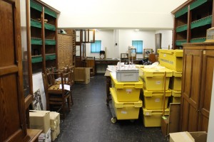
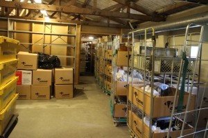
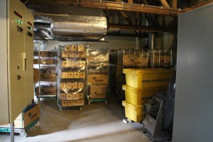
Most of the collection is being professionally moved, of course, but Prof Beijerinck’s gall and root nodule samples (preserved in alcohol) are too fragile for the vibration in a lorry and so a team of volunteers carried them around the corner to our new abode.


We plan to reopen in the Autumn, look for us at the Delft Science Centre on Mijnbouwstraat.
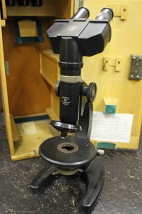
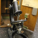
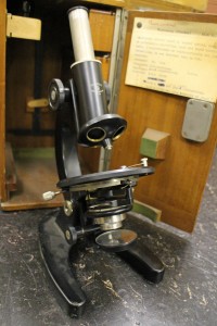
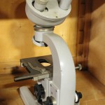
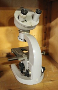
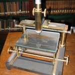
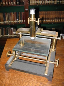
What was this microscope used for?
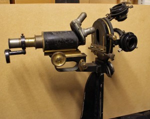
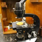
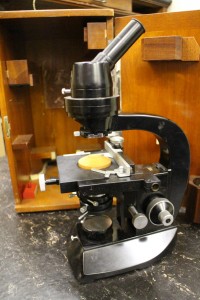
Discovering rare or unusual microscopes has become almost routine since we began packing the collection, but this one has me (and everyone else who has visited) baffled.
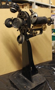
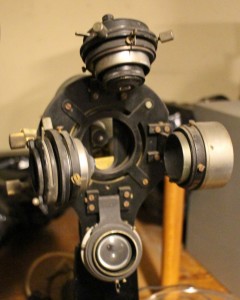
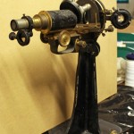
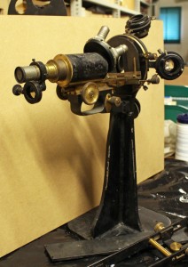
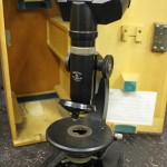
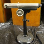
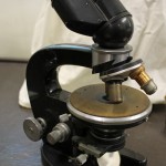
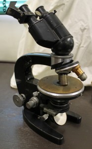
It seems to be a “jug handled” Carl Zeiss Jena microscope from the late 19th/early 20th century. However, it is mounted on its back rather than on a foot (1). Moreover, in place of the usual single condenser, there’s what seems to be 2 lenses of different strengths and 2 condensers (2), hinged so that any of them can be used to light the preparation (3). There’s an additional lens which can swing over the usual ocular (4).
Has anyone any idea what it was used for?
-
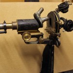
- 1: side view
-
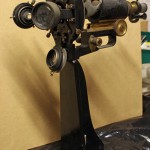
- 2: "foot"end
-
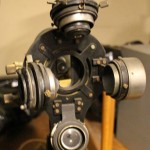
- 3: "condensers"
-
- 4: ocular end
From “out of date junk” to “exciting and rare” – our microscope collection
At the moment, sorting out the cupboards before our move has become very exciting as I’ve reached the microscope collection. Much of it was stored in the 1950s when the Laboratory of Microbiology moved from its original building, and has rarely been disturbed since then. Some of the microscopes date from the late 19th century, and even some of the 20th century ones are more interesting than might be expected.
The youngest of the companies represented in our microscope collection is probably the least well-known, especially outside the Netherlands.
Bleeker Nedoptifa
Founded by Dr Caroline E. (Lili) Bleeker and Gerard Willemse in 1939, Nedoptifa rapidly became known for the high standard of their optical products. They began with the production of binoculars for the Dutch army, but production was interrupted by WW2. Most of their microscope production seems to have been after 1945, when the “Nedoptifa” name came into use. The company cooperated with the Nobel prize-winner, Frits Zernike, in the development of phase contrast microscopy and held his patent on phase contrast microscopes. Bleeker retired at the end of 1963, the company was eventually taken over by a Delft firm and then in 1978 the factory in Zeist was closed.
Kluyver’s group seems to have used the basic Nedoptifa microscope for teaching – we have quite a few of them. Most of them have the standard circular stage, but a few are square. We also have one of their very early binocular microscopes as well as monocular and binocular phase contrast microscopes. Most of them, with the exception of the binocular phase contrast microscope, seem to have been use in in the laboratory before 1955.
Note added later: Disappointingly, after a visit in mid-June from Peter Paul de Bruyn, an expert on Bleeker microscopes, it seems that the microscopes whose boxes proclaimed them to be phase contrast microscopes do not have the necessary lenses or fittings. They may appear as we complete the packing of the collection, but it’s beginning to look unlikely.
-
- Bleeker Nedoptifa binocular microscope
-
- Early Bleeker Nedoptifa "phase contrast" microscope
-
- Bleeker "phase contrast" microscope from the 1960s
Carl Zeiss Jena
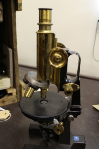
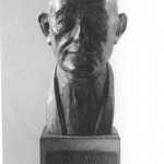
This famous old microscope manufacturer needs no introduction. In what is probably our most famous portrait of Prof Beijerinck, he is clearly using one of their “jug handled” microscopes. Others in our collection are in the Zeiss catalogues of the late 19th century.
-
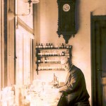
- Prof M.W. Beijerinck in his laboratory

-
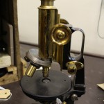
- 19th century "jug handled" Zeiss microscope
We also have some more unusual examples of their work including a “horizontal microscope” (intended for examining living plants) and a binocular microscope fitted with a prism on the right ocular to aid the drawing of samples.
-
- Horizontal Zeiss microscope

-
- Zeiss drawing microscope
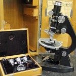
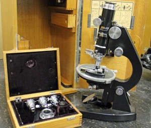
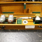
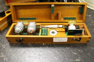
I really shouldn’t have favourites, but I must admit a fondness for the very heavy preparation microscope that turned up in a battered old wooden box on top of a cupboard. When it is taken out of the box, the eyepieces on the right flip up, giving a forerunner of our modern stereomicroscopes which can be found in the 1902 Carl Zeiss Catalogue. There are 2 sets of lenses and oculars. During dissections, the microscope can be moved backwards and forwards along the metal bar.
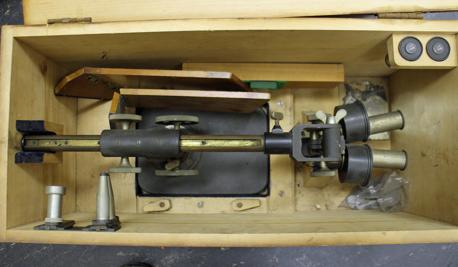
Early Zeiss preparation or dissecting microscope
Ernst Leitz Wetzlar
Another famous old company, Leitz seems to have been a favourite with Professor van Iterson. Among the microscopes from this company are a “measuring microscope”, a preparation microscope with wrist supports and the microscope (with an extensive collection of extra attachments) that he used after he retired.
-
- Leitz "measuring microscope"
-

- leitz preparation or stereo microscope

-
- Leitz microscope with one of two boxes of attachments
Other companies
Other microscope makers are mostly represented by single instruments, so I will only show two of the most spectacular. First is the interference microscope made by Cooke, Troughton & Simms from the Van Iterson collection. This microscope is accompanied by a set of extra attachments and filters.
-
- Inteference microscope by Cooke, Troughton & Simms
-
- Attachments for the Cooke et al microscope
And lastly, one of the most unusual microscopes in our collection, a reversed microscope made by Nachet & Fils of Paris. The sample lies on the stage and can be illuminated from above, with the observer’s light path underneath the stage. It appears in the 1898 Nachet catalogue. This microscope was given to the collection by Dr and Mrs ten Hoopen, both of whom worked in the Department of Applied Botany before volunteering to help with the Archive and Museum after they retired.
The Delft School of Microbiology Archive owes a huge debt to both of them. Truus undertook two major tasks. First she restored our enormous collection of glass negatives (see the blogpost “Glass negatives galore!”) after they all got damp during asbestos removal in the attic where they were stored. She then went on to research and catalogue our collections of original and printed botanic and microbiological wall charts (see blogposts “The art of Henriette Beijerinck” and “Educational Wall Charts – where are they now?”). Meanwhile, Hens catalogued the microscopes and other equipment (we have the most amazing range of pH meters, for example) in our collection.
Without Truus and Hens ten Hoopen, we would know a lot less about the Collection than we do.

Reversed light path microscope by Nachet et fils
Playing with facsimile Van Leeuwenhoek microscopes
Since most of my available time is currently vanishing into preparing the Delft School Archive and Museum for its move to Delft’s Science Centre, I thought that a simple blog showing some of the photos I’ve taken with my facsimile Van Leeuwenhoek microscopes might be nice this month.
By the way, I’ve been asked why I keep the photographs on this blog fairly small. The reason is simple – I’ve had some problems with plagiarism and this is an attempt to limit what people can do or claim with my work without the high resolution versions.
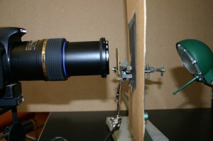
I have 5 Van Leeuwenhoek facsimiles with magnifications ranging from 68x-303x, and have been trying to repeat some of his experiments as closely as possible (it’s a good way to spend a wet weekend!). The first 2 pictures show the structure of the microscopes and my photographic setup. Ideally, one needs a camera that can cope with single point metering, and a macro lens also helps with short focusing distances. The microscope is clamped in front of the camera lens, side-on. The piece of cardboard (with a 1 cm hole in it) is there to protect the camera’s light meter because that lamp was much bigger than the microscope. I’ve recently been finding it easier, especially with the stronger lenses, to use a small LED torch.

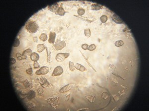
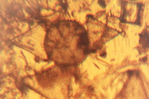
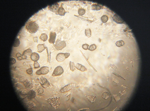
Fossil microorganisms can be very useful for comparing methods as they don’t dry out, swim away or otherwise change. Here you can see a vorticella-type protozoan (L) and diatom preparations under bright (C) and semi-dark field (R) lighting
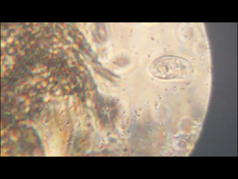
Van Leeuwenhoek’s first observation of microorganisms came from his examination of a water sample from a shallow lake called the Berkelsemeer. This lake no longer exists, but a sample from a similar lake near Delft (the Delftsehout) provided this picture (one of the first I took) of blue-green algae and a rotifer on the right. The black circle is an air bubble.

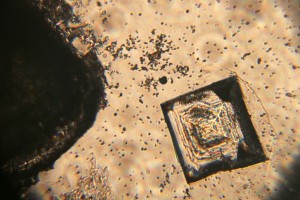
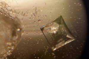
Van Leeuwenhoek did a lot of work on the formation of crystals of different sorts, and the next 2 photographs show the same table salt crystal under bright (L) and semi-dark (R) field lighting. My results agreed with Van Leeuwenhoek’s observation that you get smaller crystals if you let them form slowly by letting the saturated solution evaporate at room temperature rather than by heating it.
The next picture shows red blood cells that I’d dried onto a coverslip. I only noticed after I’d taken the photograph that the little torch had slipped a bit, but I rather like the odd lighting effect.

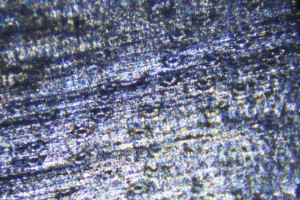
Thus far, my attempts to make thin enough slices of plant materials to examine properly have not been very successful. These photos show my best efforts for carrot root (L) and leek leaf (R). I’m reluctant to cheat by using a microtome, so will have to keep practising!

As Van Leeuwenhoek also complained, the shallow depth of field (or focus) of the stronger microscopes can be a real problem. I haven’t been very successful with taking photos of living microorganisms using the 303x lens. They tend to swim out of focus before I have time to trigger the shutter, so the least frustrating thing is to make short films and then use single frames from the film. This image shows bacteria and a hunting ciliate protozoan from a pepper water sample.
Finally, a lobster larva from a seawater sample. It was either long dead, or a shed skin as the whole thing was covered by a thin layer of algae. I had to take 2 photographs and then join them afterwards– I obviously need to get a weaker microscope!

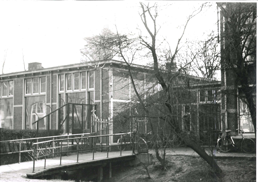
The parting of the ways…
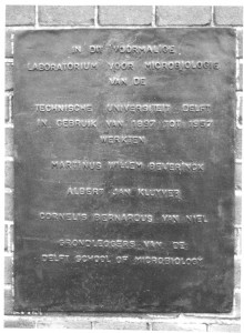
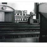
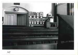
At the end of March 1958, two years after Kluyver’s death, the Laboratory for Microbiology moved out of the building where he and Beijerinck had worked. Kluyver had been heavily involved in the design and planning of the new building, but it was his successor, Torsten Wikén, who took possession.
-
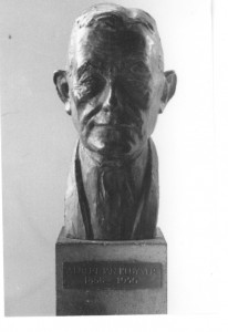
- Kluyver memorial
-
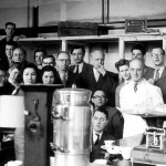
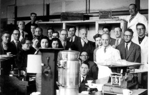
- Last day at the old lab, Prof Wiken central with a cigar
-
- Delft School memorial on old lab, Kanaalweg
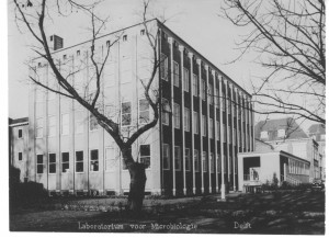
The new laboratory was attached to the new Department of Biochemistry, the Department of Applied Botany previously erected for Van Iterson and the associated Botanic Garden dedicated to applied botany.
-
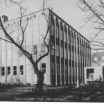
- The new Microbiology Laboratory in 1958
-
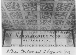
- The Beijerinck ceiling in the entrance to the Laboratory for Microbiology, 1958
Professor Wikén’s new office was given new furniture, and the contents of the office used by Beijerinck and Kluyver were stored in a purpose-built room in the microbiology attic. Since the 1980s, this collection has gradually been organised and merged with similar material left by Van Iterson when he retired and a few related donations, giving us what is known today as the Delft School of Microbiology Archives and the Museum known as the “Kamer van Beijerinck” (Beijerinck’s office). None of it would have been possible without the hard work of a legion of volunteers who have sorted and researched different areas of the collection.
The collection has attracted visitors ranging from individual researchers from as far afield as the USA and Japan to biotechnology students in their first year with their parents and visitors from schools. It’s provided material for TV programmes, exhibitions, publications and postage stamps as well as a couple of PhD theses… Visitors to the Department of Biotechnology have frequently been brought to see the collection during their visits, as have participants in some of the Delft Advanced Courses.
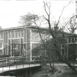
All good things eventually come to an end, and it is finally time for the Archive-Museum and the Department of Biotechnology to part company. Biotechnology is moving to a brand new Faculty building on the outskirts of Delft, uniting with other Faculty Departments. The Archive-Museum is moving around the corner to the old Department of Mining building, now the Delft Science Centre where the collection will occupy second floor rooms over the main entrance, next door to the minerals collection of the Department of Mining.
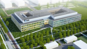
-

- Artist's impression of the new Faculty building
-
- Delft Science Centre with new Kamer van Beijerinck, 2nd floor with balcony

Regular readers of this blog will know that digitisation and cataloguing of the collection has been an on-going process and we’ve frequently been surprised as volunteers have found manuscripts and odd equipment as they’ve catalogued the contents of boxes. As we prepare for the move this is still true, and readers will no doubt be hearing about some of our more unusual discoveries (e.g. the papers relating to the unmasking of a cold war spy in the Department) in later posts.
Saying farewell to the Julianalaan will be sad in some ways (most of my scientific career was spent there), but our new rooms will be better lit and considerably less dusty. The Museum will be more accessible as it will no longer be necessary to walk through an active biotechnological laboratory to reach it. Last and not least, the move is giving us a chance to sort the document collection more logically, something that researchers who’ve visited us in search of specific documents will appreciate!
Delft’s Biological Labs 2: The originals..
Sadly, there are no pictures of the first “microbiological laboratory” and the building was demolished at the start of the 1890s, but by collecting the occasional descriptive comments from Antoni van Leeuwenhoek’s letters, we can gain a glimpse of the room where so many types of microorganism were seen for the first time. The following is an extract from our book “Antoni van Leeuwenhoek: Master of the Miniscule” which is scheduled for publication by Brill, Leiden (Brill.nl) in April, 2016.
Van Leeuwenhoek’s home and workshop
Antoni van Leeuwenhoek worked in his comptoir. The word literally means a counting or writing room, but it was actually his workshop. It was located in an annex to his house so that he would not be disturbed. The room was partitioned off with wainscoting, with a hole to accommodate the spring pole of his lathe. The bottom two of the four windows overlooking the street could be opened upwards, and were fitted with wooden shutters. His workshop could be closed off “so that little or no air enters from outside”, but if he was working with a candle, he preferred to open them a little, and cover the window with curtains.
His letters and the inventory of the house compiled after the death of his daughter Maria reveal some of the equipment and materials that he used. As well as his lens and microscope making equipment, there was a small furnace to extract gold and silver from ore, a barometer, a salt water aquarium, sharp blades, chemicals (including the distillate “brandewijn” for conservation and saffron for colouring), and a collection of specimens ranging from minerals to the testicles of a rat preserved in spirits. He had a balance with weights, a glass-blowing table with a lamp, and an anvil for the production of his microscopes, and all of his surveying equipment.
Many of the objects that Van Leeuwenhoek studied came from the immediate surroundings of his home. He had two gardens, one adjoining the house with a well and another outside the city. He kept a green parrot for a long time and examined the excrement of the sparrows in his yard after feeding them. Delft’s fish market was just across the canal from his house. Since he regularly attended the dissection demonstrations at the neighbouring Anatomy Theatre, it seems likely that he brought samples home. Additionally, in a few of his letters he mentioned that sailors returning from voyages brought him exotic samples.

The star shows the position of Van Leeuwenhoek’s house in relation to the Town Hall, where he worked, the marketplace and the Old Church where he is buried.
The Microbiology Laboratory
Beijerinck originally came to Delft in 1885 to set up and run a microbiological laboratory in J.C. van Marken’s Yeast Factory. Van Marken and his wife were enlightened employers, providing their workers and their families with on-site houses, education, medical and social facilities. Van Marken made it a point of honour to keep all of his staff informed about the factory by means of an internal paper called “De Fabrieksbode”. In 1885 he wrote an article about the need for proper bacteriological studies, edited extracts of which follow:
“I would like to explain the need for the appointment of Dr. Mr. W. Beijerink as Chief of the Bacteriological Laboratory of the Yeast Factory…..
I have previously discussed the idea that “life is a struggle”, in this case between yeast and against harmful bacteria. The day on which the last harmful bacteria has been expelled from our
factory will be a cause for celebration and a public holiday….
We would have a quality supply of yeast, which would overcome the sharpest competition; our yeast would be able to circle the world in 80 days, without spoiling.
So fight the harmful bacteria!“
Van Marken’s attitude to results seems remarkably relaxed, contrasting with the attitudes of modern industry. He went on to say:
”Whatever the case, the arrival of the scholar, Dr Beyerinck must be appreciated in more ways than one. We must not have exaggerated expectations of his activities in and for our factory, but have faith that in maybe one, five or even ten years, some day a ray of light will be cast into the darkness of the fermentation business and bring incalculable advantages to our company.”
It cannot be claimed that Beijerinck was happy as an industrial scientist. He felt responsible if errors incurred financial losses, and his interests were much wider than required by his job. As can be seen from the table below and his laboratory journals, he seems to have normally had several unrelated lines of research running at any one time. Only the rows marked * involved work needed by the Factory.
TOPICS OF PUBLICATIONS THAT APPEARED DURING BEIJERINCK’S INDUSTRIAL YEARS
Sunsets (Were the spectacular sunsets of the time due to dust from Krakatoa?)
Root nodules and their bacteria
Plant galls
Grasses, carrots, gardenias, barley
Algae, protozoa in drinking water, hydrogen peroxide in living organisms
*Fermentation, butanol fermentation, Saccharomyces associated with beer, Schizosaccharomyces octosporus
Lactase, maltase, blue cheese bacteria, kefir
Photobacteria, sulfate reduction
*Methods: auxanograms, gelatine plates, Chamberland filters, sampling stratified cultures, microbiochemical analysis.
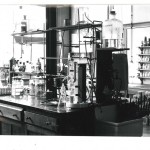
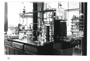
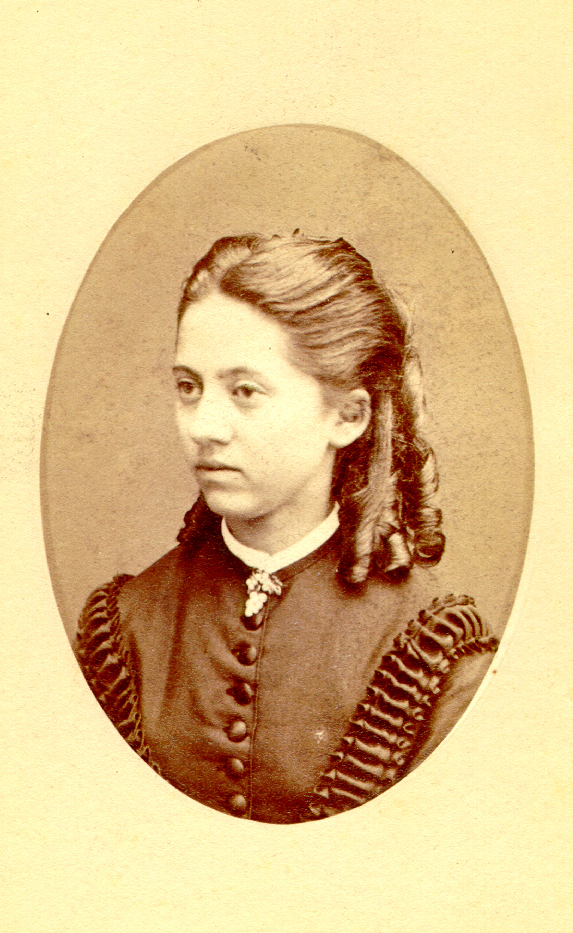
When his sisters, Henriette and Johanna, visited him in his laboratory, Henriette reported that he “sat there, surrounded by a mass of retorts, bottles and glasses, boxes, corks and heating apparatus, so that it looked like the workshop of an alchemist”.

Eventually, Van Marken and a few others managed to persuade the Delft Polytechnic (now Delft University of Technology) that they really needed a Department of Microbiology with Beijerinck as its Professor. Until his new lab was ready, he continued to use the laboratory at the Yeast Factory. Their support continued for the rest of his life – when Beijerinck retired, the Yeast Factory paid for the construction of a laboratory in the garden of his retirement house in Gorssel.
Applied Botany
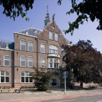
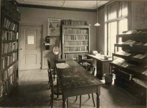
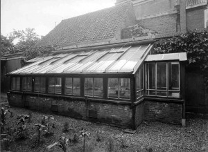
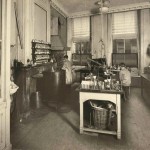
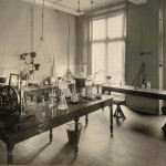
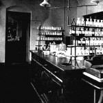
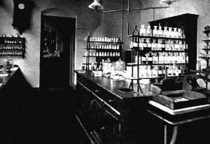
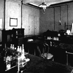
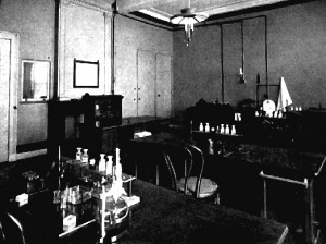
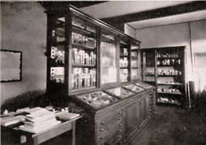
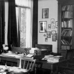
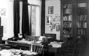
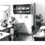
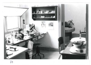
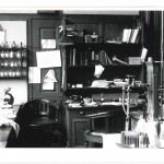
After his appointment as a Professor, Van Iterson initially worked in Beijerinck’s lab , but in 1908 he was given space in a house at Oude Delft 81, a building used for temporary accommodation by the Polytechnic. As can be seen from the photographs, the rooms were small and not really suitable for use as teaching laboratories. The office, and the library were tiny, and the greenhouse was far too small for a Department called “Applied Botany”. It wasn’t until 1911, when he was offered a job in Java, that the Polytechnic agreed to start work on a purpose-built laboratory and Botanic garden. Both finally opened in 1917. At the time of writing, the laboratory is now part of Delft University’s Department of Biotechnology, but the Department will move to a new building later this year. The Garden’s website is HERE.
-
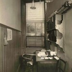
- Office
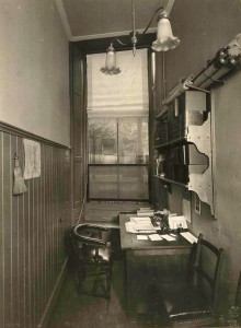
-
- Library
-

- Greenhouse
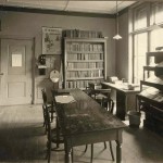
-
- Laboratory

-
- Research laboratory

-
- Teaching lab
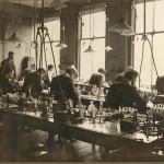
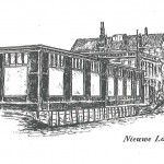
Delft University’s Biological Labs 1: The “Palace of Light” on the Nieuwelaan.
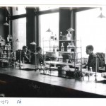
At the beginning of 2016, the Laboratory for Microbiology, together with the rest of the Department of Biotechnology, will move out of its current building, destination the new Faulty building on the outskirts of Delft. This seems like a good moment to take a look at the previous lab where so many breakthroughs were made. It was known as the “Palace of Light” because people worked late into the night.
We are fortunate that, thanks to a commemorative book published when Beijerinck was a fairly new Professor and photographs taken just before the department moved out, we know what it looked like. At the time it was built, it was the most expensive laboratory in the University. An extension was added in 1911.
Beijerinck’s laboratory
-
- The 1911 extension

-
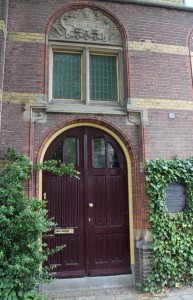
- The new door into the lab

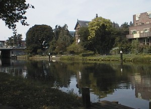
Part of the building, on the left of the top photo, was the Professor’s house. The garden sloped gently down to a large canal (the canal’s edge can be seen as a white line across the bottom of the picture). It was in the greenhouse in this garden that the tobacco plants were grown for Beijerinck’s work on the Tobacco Mosaic Virus, and many famous microbial species were isolated from the soil and mud of the garden and canal.
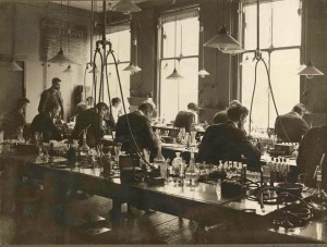
We have a few pictures of the inside of the lab in Beijerinck’s time.
-
- staff lab
-
- student lab
-
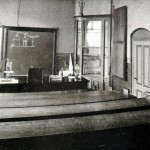
- lecture theatre
-
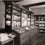
- culture collection
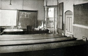
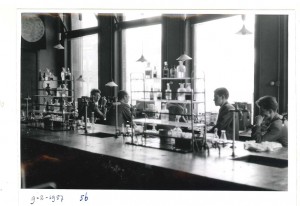
After Kluyver’s death and before everybody moved to the new (and current) building, everything was photographed. The photos are all numbered with corresponding floor plans so that we know not only which room is shown, but where the photographer was standing to take the picture. This is just a small sample.
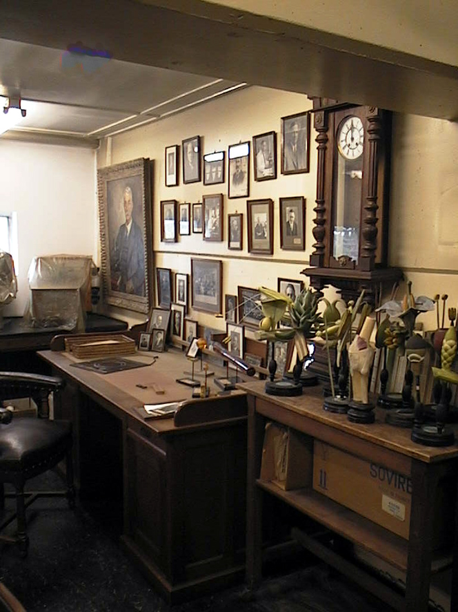
-
- Kluyver's office as he left it.
-
- Kluyver's lecture theatre
-
- Electron microscopy
-
- Staff lab

-
- Students laboratory
-
- Laboratory
The building (without the extension) is still there. After a number of trials and tribulations including the building of a major bridge close to the front door of Beijerinck and Kluyver’s house, it is now apartments. To mark the 100th anniversary of Beijerinck’s appointment, a plaque was unveiled next to the original laboratory door.

The path to a ground-breaking paper
Martinus Beijerinck started his career as a botanist. His Doctor’s thesis was about plant galls and the insects that cause them, a subject that obviously held his attention since he continued to collect galls throught his time as a Professor of Microbiology.
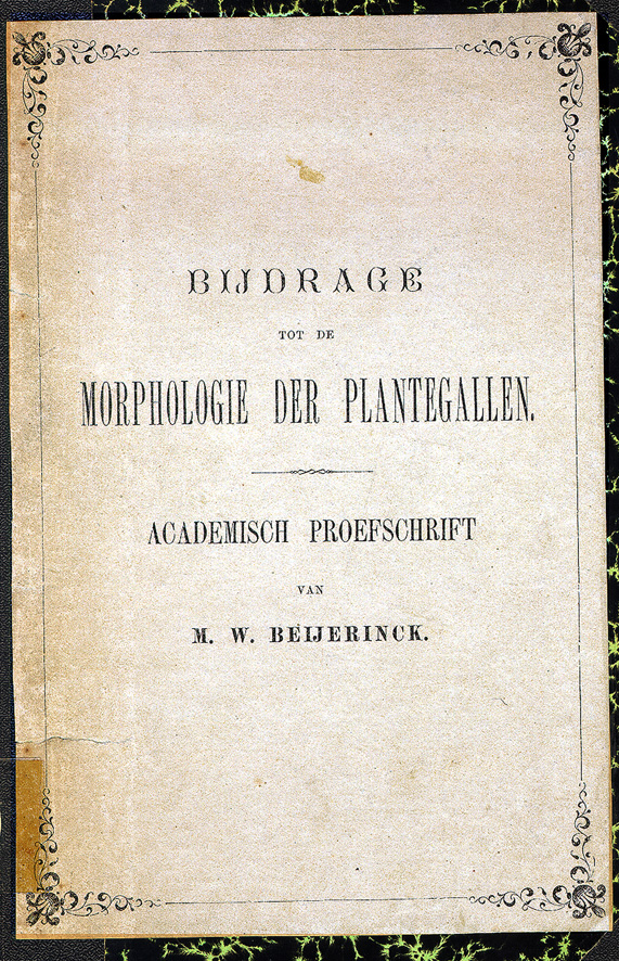
Beijerinck’s thesis
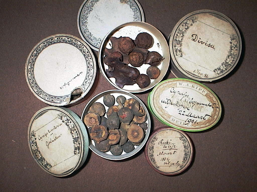
Dried galls from the Beijerinck collection
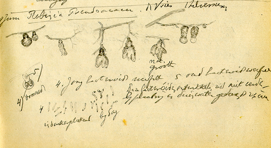
In his thesis, Beijerinck wrote that he was frustrated by his inability to find the gall wasp that was causing galls on the roots of some species of plant, notably the pea family. These nodules had different shapes, depending on the sort of plant.
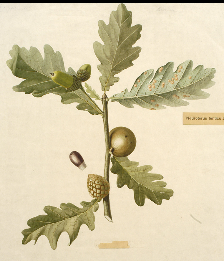
Oak apple galls by Henriette Beijerinck
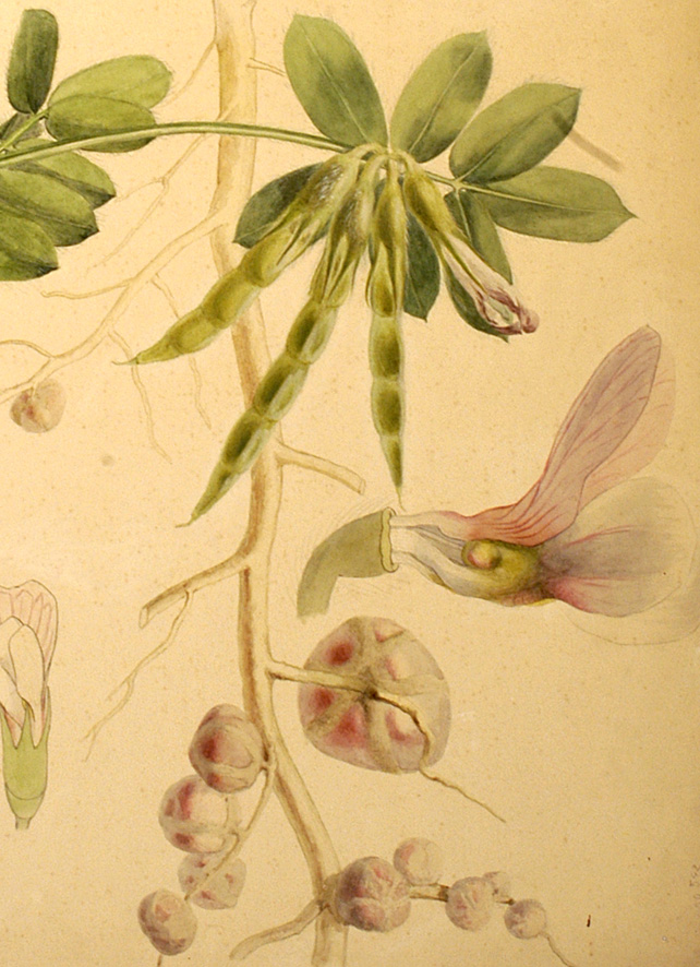
Vicia faba nodules by Henriette Beijerinck
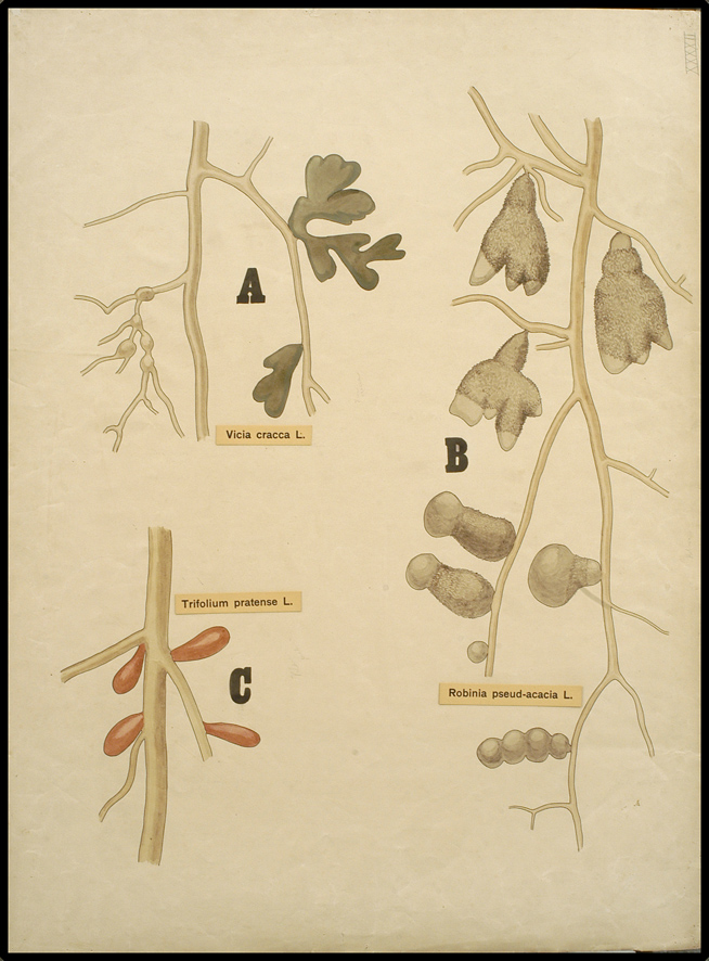
Root nodules from different species by Henriette Beijerinck
It is easy to speculate that it was this subject that sparked his interest in microbiology because when he crushed the nodules and examined the resulting tissue under the microscope, it was clear to see that a lot of bacteria were present. Some of them had an unusual Y shape. Winogradsky had recently published about nitrogen-fixing bacteria, and when Beijerinck tested his new bacteria, he found that they could also take nitrogen from the air and make ammonium.
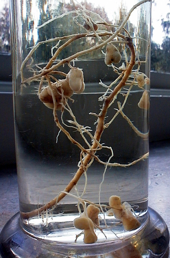
Beijerinck’s root nodules in alcohol
Root nodules and the bacteroids from Beijerinck’s lab journal
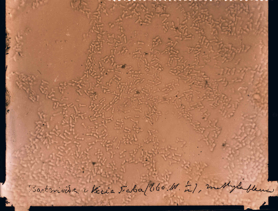
Rhizobium species under the light microscope
The first nodules that he studied (in 1888) came from the broad bean (Vicia faba), and the bacteria are now known to be a species of Rhizobium. Nitrogen fixing bacteria are responsible for maintaining soil fertility, and gardeners have known for centuries that it is a good idea to rotate crops, growing peas and beans with their root nodules to restore soil that has been used for other plants. This is why these plants are known to many as “green fertiliser”.
The art of Henriette Beijerinck
The laboratory was well supplied with printed botanic wall charts, but there was always a need for things that weren’t available. During Beijerinck’s professorship, his sister Henriette provided most of his display material, generally as large (A1 or A2 in modern terms) watercolours. Henriette Beijerinck was a qualified art teacher who presumably had private pupils, and in addition to material to illustrate lectures, also provided illustrations for several books.
-
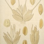
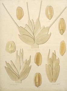
- agricultural grains
-
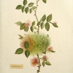
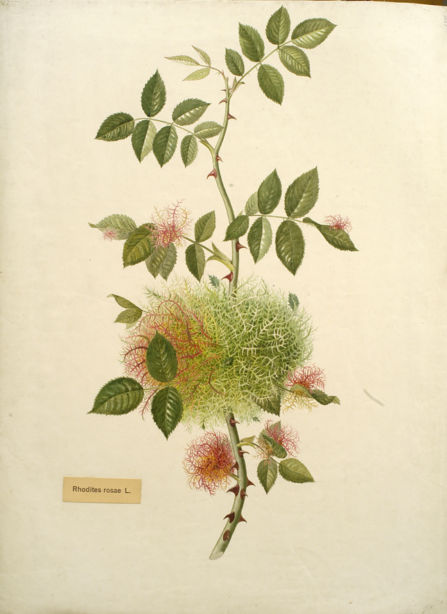
- rose gall
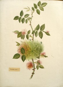
Professor Beijerinck’s earliest publications (while he was working at the Wageningen Agricultural Collece) were about the improvement of grains for agriculture, and our collection contains a number of drawings of seedheads. Since they are not signed, they could be by Henrietta, or even by Martinus. The image above left shows (clockwise from bottom left) wheat, European spelt and duram wheat. Beijerinck’s doctoral thesis (and a lifelong subject of research) was about plant galls and the collection includes quite a few pictures of galls. The illustration above right shows moss galls caused by Rhodites rosa gall wasp on young rose leaves.
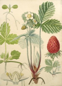
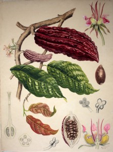
Henriette also painted assorted microorganisms (see, for examples, the blog post about possible life in comets), but her most beautiful works are the botanic charts. These three images are among the best.
-
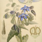
- Borage

-
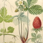
- Strawberry
-
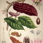
- Cocoa
Finding more van Leeuwenhoek microscopes…
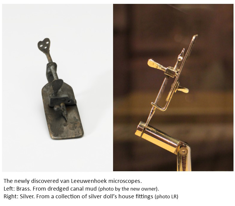
In April, I published a survey of the currently accepted authentic Van Leeuwenhoek microscopes in FEMS Microbiology Letters, and suggested that more might be found. Within a month, there came news of two previously unknown microscopes. Their history over the intervening centuries could not have been more different.
Press releases in the Dutch newspapers and on TV (21st May, 2015) announced that a silver Van Leeuwenhoek microscope had been discovered in a box of old Dutch doll’s house equipment. The new owner took it to the Museum Boerhaave in Leiden for authentication. They compared it with the 4 Van Leeuwenhoek microscopes already in their collection and the microscope belonging to the University of Utrecht before presenting their results at the beginning of June. They concluded that this microscope indeed dates from Van Leeuwenhoek’s lifetime and must be considered to be authentic. It can be seen at Museum Boerhaave until mid-July (2015). Thereafter it will go home to its owner for display at Planetarium Zuylenburgh in Oud-Zuilen.
Within days of the announcements about the silver microscope, a letter by Brian Ford appeared in the journal “Nature” (28 May, 2015), announcing the finding of a brass Van Leeuwenhoek microscope in sediment that had been dredged from Delft canals during maintenance work. This microscope’s new owner had sent it for authentication to Dr Ford in the UK, and he had concluded that it was indeed real.
Before these two discoveries, the most recent find was in 1983, when a visitor to the “Beads of Glass” exhibition at Museum Boerhaave donated a small silver microscope that she had owned without realizing what it was.
It is likely that many of Van Leeuwenhoek’s gold and silver microscopes have been melted down, but my crystal ball tells me that there must be more out there to find, especially since he also made a number from brass. For example, two microscopes were individually photographed at the start of the 20th century in Munich and Paris, but their locations are now unknown. From the photograph in the 1929 Nachet Collection Catalogue, it can be seen that the Paris microscope had been incorrectly assembled and could not have been used until it had been put together properly, a mistake that someone making a copy would be unlikely to make. The Munich microscope had been accepted as authentic by the University of Utrecht in the late 19th century, before it was sent to Munich.

Like the newly discovered microscopes, there are structural similarities, but neither appears to be physically identical to the accepted microscopes. This rules out most of the known late 19th and 20th century copies (which tend to be replicas of either the Utrecht University microscope or one from the Museum Boerhaave collection). Hopefully they (and others) will reappear, and can undergo modern authentication analysis.
Recent Comments