
Posts tagged microbiology
Playing with facsimile Van Leeuwenhoek microscopes
Since most of my available time is currently vanishing into preparing the Delft School Archive and Museum for its move to Delft’s Science Centre, I thought that a simple blog showing some of the photos I’ve taken with my facsimile Van Leeuwenhoek microscopes might be nice this month.
By the way, I’ve been asked why I keep the photographs on this blog fairly small. The reason is simple – I’ve had some problems with plagiarism and this is an attempt to limit what people can do or claim with my work without the high resolution versions.
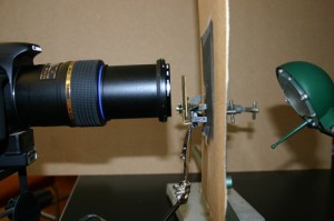
I have 5 Van Leeuwenhoek facsimiles with magnifications ranging from 68x-303x, and have been trying to repeat some of his experiments as closely as possible (it’s a good way to spend a wet weekend!). The first 2 pictures show the structure of the microscopes and my photographic setup. Ideally, one needs a camera that can cope with single point metering, and a macro lens also helps with short focusing distances. The microscope is clamped in front of the camera lens, side-on. The piece of cardboard (with a 1 cm hole in it) is there to protect the camera’s light meter because that lamp was much bigger than the microscope. I’ve recently been finding it easier, especially with the stronger lenses, to use a small LED torch.

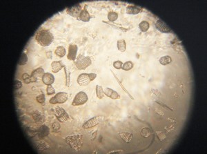
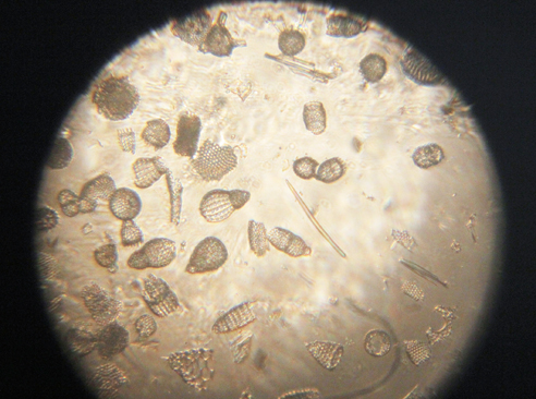
Fossil microorganisms can be very useful for comparing methods as they don’t dry out, swim away or otherwise change. Here you can see a vorticella-type protozoan (L) and diatom preparations under bright (C) and semi-dark field (R) lighting
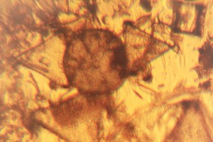
Van Leeuwenhoek’s first observation of microorganisms came from his examination of a water sample from a shallow lake called the Berkelsemeer. This lake no longer exists, but a sample from a similar lake near Delft (the Delftsehout) provided this picture (one of the first I took) of blue-green algae and a rotifer on the right. The black circle is an air bubble.

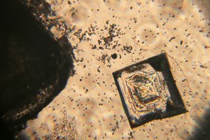
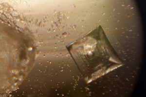
Van Leeuwenhoek did a lot of work on the formation of crystals of different sorts, and the next 2 photographs show the same table salt crystal under bright (L) and semi-dark (R) field lighting. My results agreed with Van Leeuwenhoek’s observation that you get smaller crystals if you let them form slowly by letting the saturated solution evaporate at room temperature rather than by heating it.
The next picture shows red blood cells that I’d dried onto a coverslip. I only noticed after I’d taken the photograph that the little torch had slipped a bit, but I rather like the odd lighting effect.

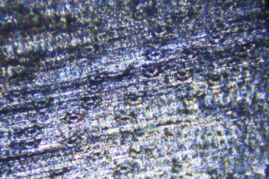
Thus far, my attempts to make thin enough slices of plant materials to examine properly have not been very successful. These photos show my best efforts for carrot root (L) and leek leaf (R). I’m reluctant to cheat by using a microtome, so will have to keep practising!

As Van Leeuwenhoek also complained, the shallow depth of field (or focus) of the stronger microscopes can be a real problem. I haven’t been very successful with taking photos of living microorganisms using the 303x lens. They tend to swim out of focus before I have time to trigger the shutter, so the least frustrating thing is to make short films and then use single frames from the film. This image shows bacteria and a hunting ciliate protozoan from a pepper water sample.
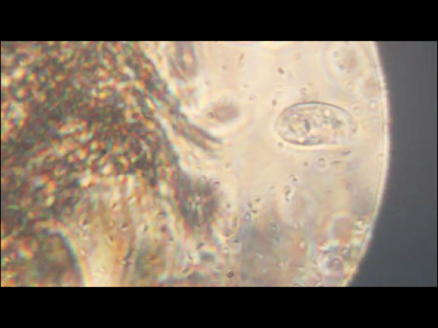
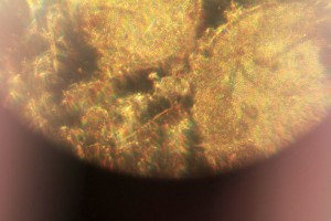
Finally, a lobster larva from a seawater sample. It was either long dead, or a shed skin as the whole thing was covered by a thin layer of algae. I had to take 2 photographs and then join them afterwards– I obviously need to get a weaker microscope!

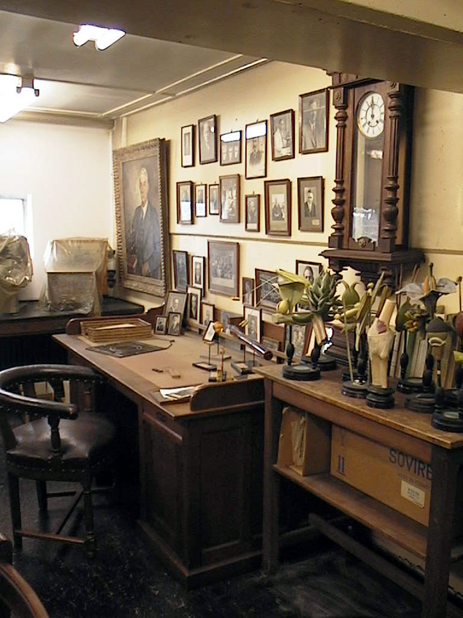
The Delft School of Microbiology Archives
The “Delft School of Microbiology” is based on the work of our first three biological Professors – Beijerinck, van Iterson and Kluyver. The Archive is a collection of their papers, teaching aids, furniture and other materials and functions as a small museum in the Department of Biotechnology.
At the moment, the collection is being digitised and catalogued. This blog will report on some of the interesting and sometimes unexpected things to be found in the collection.
We’ll also cast an occaisional glance at our honorary member, the original Delft microscopist and microbiologist, Antonie van Leeuwenhoek.
Recent Comments