
Posts tagged simple microscopes
Van Musschenbroek Microscopes
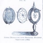
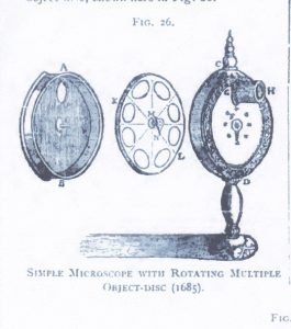
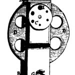
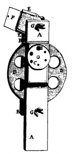
It cannot be denied that Antoni van Leeuwenhoek made major discoveries with his single lensed microscopes. Consequently, when thinking about the simple microscopes of the 16th and 17th centuries, most people have focused on his very simple (some say crude) microscopes. In fact, there were several other single lensed microscopes in use at the time. Many of them were more complicated than Van Leeuwenhoek’s microscope with extra features and even engraved ornamentation. One had a rotating disc with different sized holes to give varying apertures, others had detachable lenses or moving sample holders so that samples did not have to be transferred to another microscope for a change of magnification. Significant discoveries were also made with these microscopes and yet they are rarely studied. Comparison of results from the different types could tell us so much more, but working replicas (with proper lenses) like the Van Leeuwenhoek replicas I normally work with are not to be found.
-
- Kircher
-
- Huygens 1
-
- Huygens ver 2
-
- Joblot
-
- Hartsoeker

-
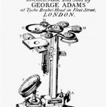
- Adams

Last year, after I complained about the lack of such replicas for experimental use during a lecture, someone called my bluff. Dr Koen Quint of the Dutch Stichting voor Historische Microscopie offered me the honour of trying their (real) Van Musschenbroek microscope! I have to admit that my first reaction was panic.
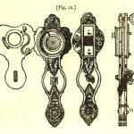
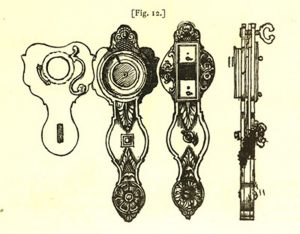
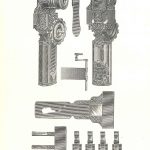
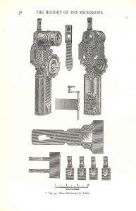
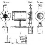
The Van Musschenbroek workshop in Leiden was a family business between 1660 and 1750. They produced (and invented) a range of scientific instruments, among which were microscopes. At its peak between 1730 and 1740, their microscope range included 10 different models, some of which were their own invention. The more complicated of their single lensed microscopes included a holder and detachable lenses.

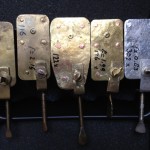
Van Musschenbroek microscope with lenses and sample holders
The initial experiments have been encouraging. Of course, using a centuries-old microscope was a bit nerve wracking, and much of the first session was spent working out how to set everything up without damaging it (e.g. we wrapped the handle of the microscope in felt to protect it against the clamps holding it erect). Obviously I can’t take it home to use on wet Sunday afternoons as I would with a replica, but the next few weeks are going to be fun!

Experimental setup
Playing with facsimile Van Leeuwenhoek microscopes
Since most of my available time is currently vanishing into preparing the Delft School Archive and Museum for its move to Delft’s Science Centre, I thought that a simple blog showing some of the photos I’ve taken with my facsimile Van Leeuwenhoek microscopes might be nice this month.
By the way, I’ve been asked why I keep the photographs on this blog fairly small. The reason is simple – I’ve had some problems with plagiarism and this is an attempt to limit what people can do or claim with my work without the high resolution versions.
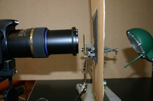
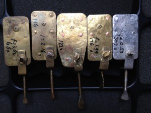
I have 5 Van Leeuwenhoek facsimiles with magnifications ranging from 68x-303x, and have been trying to repeat some of his experiments as closely as possible (it’s a good way to spend a wet weekend!). The first 2 pictures show the structure of the microscopes and my photographic setup. Ideally, one needs a camera that can cope with single point metering, and a macro lens also helps with short focusing distances. The microscope is clamped in front of the camera lens, side-on. The piece of cardboard (with a 1 cm hole in it) is there to protect the camera’s light meter because that lamp was much bigger than the microscope. I’ve recently been finding it easier, especially with the stronger lenses, to use a small LED torch.

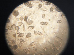
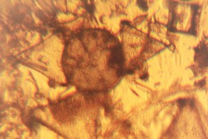
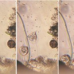
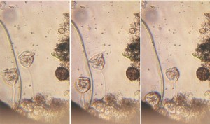
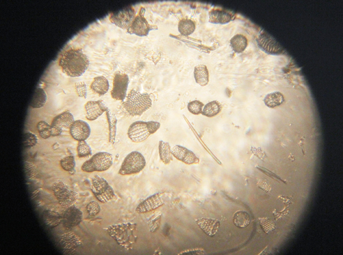
Fossil microorganisms can be very useful for comparing methods as they don’t dry out, swim away or otherwise change. Here you can see a vorticella-type protozoan (L) and diatom preparations under bright (C) and semi-dark field (R) lighting
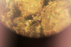
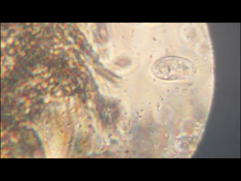
Van Leeuwenhoek’s first observation of microorganisms came from his examination of a water sample from a shallow lake called the Berkelsemeer. This lake no longer exists, but a sample from a similar lake near Delft (the Delftsehout) provided this picture (one of the first I took) of blue-green algae and a rotifer on the right. The black circle is an air bubble.

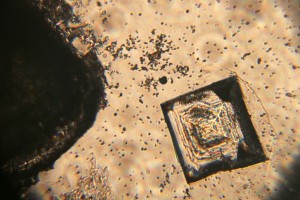
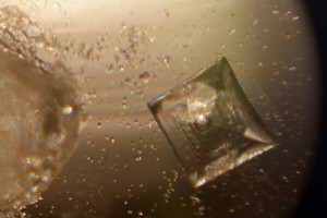
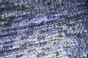
Van Leeuwenhoek did a lot of work on the formation of crystals of different sorts, and the next 2 photographs show the same table salt crystal under bright (L) and semi-dark (R) field lighting. My results agreed with Van Leeuwenhoek’s observation that you get smaller crystals if you let them form slowly by letting the saturated solution evaporate at room temperature rather than by heating it.
The next picture shows red blood cells that I’d dried onto a coverslip. I only noticed after I’d taken the photograph that the little torch had slipped a bit, but I rather like the odd lighting effect.

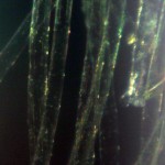
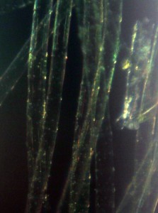
Thus far, my attempts to make thin enough slices of plant materials to examine properly have not been very successful. These photos show my best efforts for carrot root (L) and leek leaf (R). I’m reluctant to cheat by using a microtome, so will have to keep practising!

As Van Leeuwenhoek also complained, the shallow depth of field (or focus) of the stronger microscopes can be a real problem. I haven’t been very successful with taking photos of living microorganisms using the 303x lens. They tend to swim out of focus before I have time to trigger the shutter, so the least frustrating thing is to make short films and then use single frames from the film. This image shows bacteria and a hunting ciliate protozoan from a pepper water sample.
Finally, a lobster larva from a seawater sample. It was either long dead, or a shed skin as the whole thing was covered by a thin layer of algae. I had to take 2 photographs and then join them afterwards– I obviously need to get a weaker microscope!

Finding more van Leeuwenhoek microscopes…
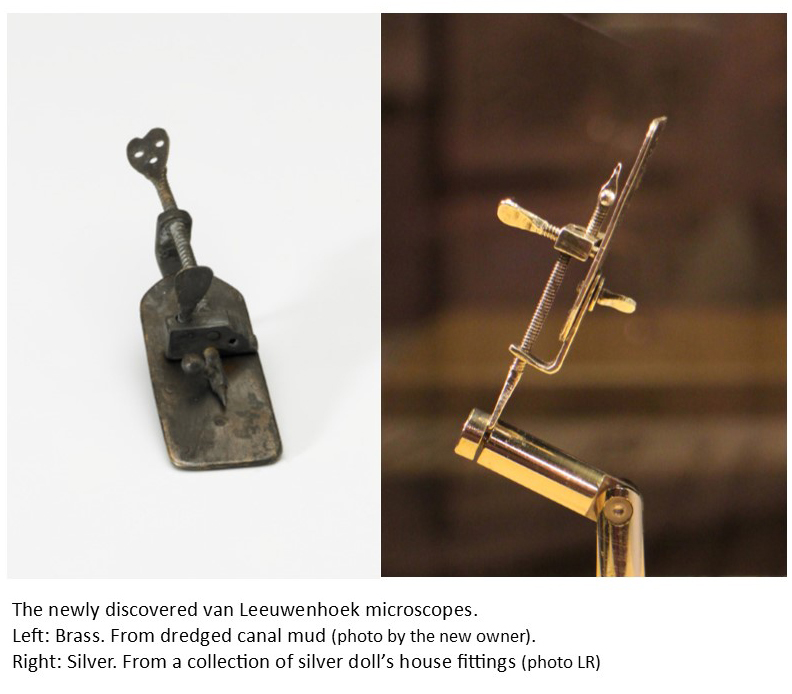
In April, I published a survey of the currently accepted authentic Van Leeuwenhoek microscopes in FEMS Microbiology Letters, and suggested that more might be found. Within a month, there came news of two previously unknown microscopes. Their history over the intervening centuries could not have been more different.
Press releases in the Dutch newspapers and on TV (21st May, 2015) announced that a silver Van Leeuwenhoek microscope had been discovered in a box of old Dutch doll’s house equipment. The new owner took it to the Museum Boerhaave in Leiden for authentication. They compared it with the 4 Van Leeuwenhoek microscopes already in their collection and the microscope belonging to the University of Utrecht before presenting their results at the beginning of June. They concluded that this microscope indeed dates from Van Leeuwenhoek’s lifetime and must be considered to be authentic. It can be seen at Museum Boerhaave until mid-July (2015). Thereafter it will go home to its owner for display at Planetarium Zuylenburgh in Oud-Zuilen.
Within days of the announcements about the silver microscope, a letter by Brian Ford appeared in the journal “Nature” (28 May, 2015), announcing the finding of a brass Van Leeuwenhoek microscope in sediment that had been dredged from Delft canals during maintenance work. This microscope’s new owner had sent it for authentication to Dr Ford in the UK, and he had concluded that it was indeed real.
Before these two discoveries, the most recent find was in 1983, when a visitor to the “Beads of Glass” exhibition at Museum Boerhaave donated a small silver microscope that she had owned without realizing what it was.
It is likely that many of Van Leeuwenhoek’s gold and silver microscopes have been melted down, but my crystal ball tells me that there must be more out there to find, especially since he also made a number from brass. For example, two microscopes were individually photographed at the start of the 20th century in Munich and Paris, but their locations are now unknown. From the photograph in the 1929 Nachet Collection Catalogue, it can be seen that the Paris microscope had been incorrectly assembled and could not have been used until it had been put together properly, a mistake that someone making a copy would be unlikely to make. The Munich microscope had been accepted as authentic by the University of Utrecht in the late 19th century, before it was sent to Munich.

Like the newly discovered microscopes, there are structural similarities, but neither appears to be physically identical to the accepted microscopes. This rules out most of the known late 19th and 20th century copies (which tend to be replicas of either the Utrecht University microscope or one from the Museum Boerhaave collection). Hopefully they (and others) will reappear, and can undergo modern authentication analysis.
Delft’s first microbiologist – Antonie van Leeuwenhoek
Although the Delft School of Microbiology only dates back to Martinus Beijerinck and the late 19th century, it seems churlish to ignore Antonie van Leeuwenhoek on a blog discussing Delft microbiology just because he was 200 years too early. He was not a teacher and indeed actively resisted explaining his methods, but he did publish copiously about everything he saw with his magnifying glasses and simple microscopes, making him the first microbiologist (although not the first microscopist).
Today, van Leeuwenhoek is generally mentioned in connection with the discovery of microorganisms. However, his studies were much broader than that. He dissected insects, and examined anything that would fit on his microscope. His first letter to the Royal Society illustrates this clearly as it covers the sting, head and eye of the bee, and the structure of a louse as well as his observations of fungus that he said grew on leather, meat and other things.
Van Leeuwenhoek’s microbiological discoveries began in 1674 when he examined samples from the cloudy water of the Berkelsemeer, a lake near Delft that no longer exists, and found his famous “little animals”. His discovery of bacteria probably dates from his pepper water experiments in 1676, when he reported seeing extremely small animals among the others – a copy of the drawing that accompanied this letter was published by Henry Baker, and is shown here. “Fig IV” is probably the first appearance in print of a bacterium.
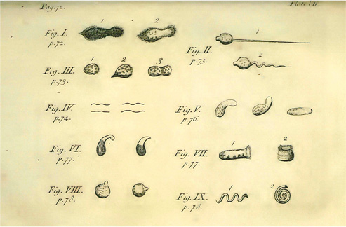
Baker’s copy of AvL’s pepper water illustration.
-
- Bright field microscopy of living protozoa
-
- Dark field microscopy of algae
-
- Facsimiles of van Leeuwenhoek microscopes
The film clip here – www.youtube.com/watch?v=OniSF8QrHac – shows what can be seen with facsimiles of van Leeuwenhoek microscopes.
And there’s an excellent website about our Founding Father here: http://lensonleeuwenhoek.net/
Recent Comments