
Delft Microbiology
Thoughts on Van Leeuwenhoek’s optical tool box
My personal project to find the descriptions of methods buried in Van Leeuwenhoek’s letters and to try and work out how he did things that he didn’t describe continues. My most recent results have now appeared in the Nov 2017 FEMS Letters with the POI: https://doi.org/10.1093/femsle/fnx247. The paper covers problems encountered with using the classical microscopes with some samples that we know he studied, and suggests a solution.
Antoni van Leeuwenhoek did not limit his research to microorganisms. Indeed, he worked with a very wide range of samples ranging from wood and animal tissues through insects to individual cells and sperm to bacteria. His microscopes work very well with transparent samples where the light can pass through them (and the samples were small) but, as can be seen from Figure 1, he was obviously also using reflected light.
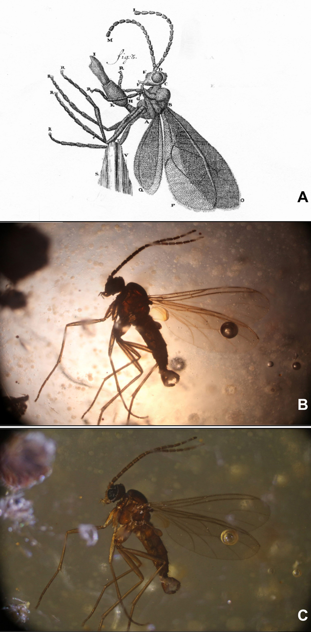
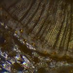
Transmitted and reflected lighting of a gnat
Figure 1A shows a drawing of a gnat, taken from a 1702 Van Leeuwenhoek letter. Figures 1B and C. show a similar insect photographed under a 19th century Carl Zeiss Jena microscope with transmitted (B) and reflected (C) light. It is clear that the level of detail shown by Van Leeuwenhoek cannot be observed on the silhouette shown in B.
The details are all in my papers, but my conclusion is that, as he did with his aalkijkers (his microscopes for viewing the blood in the capillaries of the tails of small eels and fish where the sample pin was replaced by a glass tube to keep the specimen alive), Van Leeuwenhoek adapted his magnifiers to suit the task in hand.
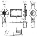
Figure 2 shows a collection of magnifying equipment that I think Van Leeuwenhoek used as his optical tookbox. The photographs show modern facsimiles, the drawings were made during or just after Van Leeuwenhoek’s life. This conclusion is based on experimental results and historical documents.
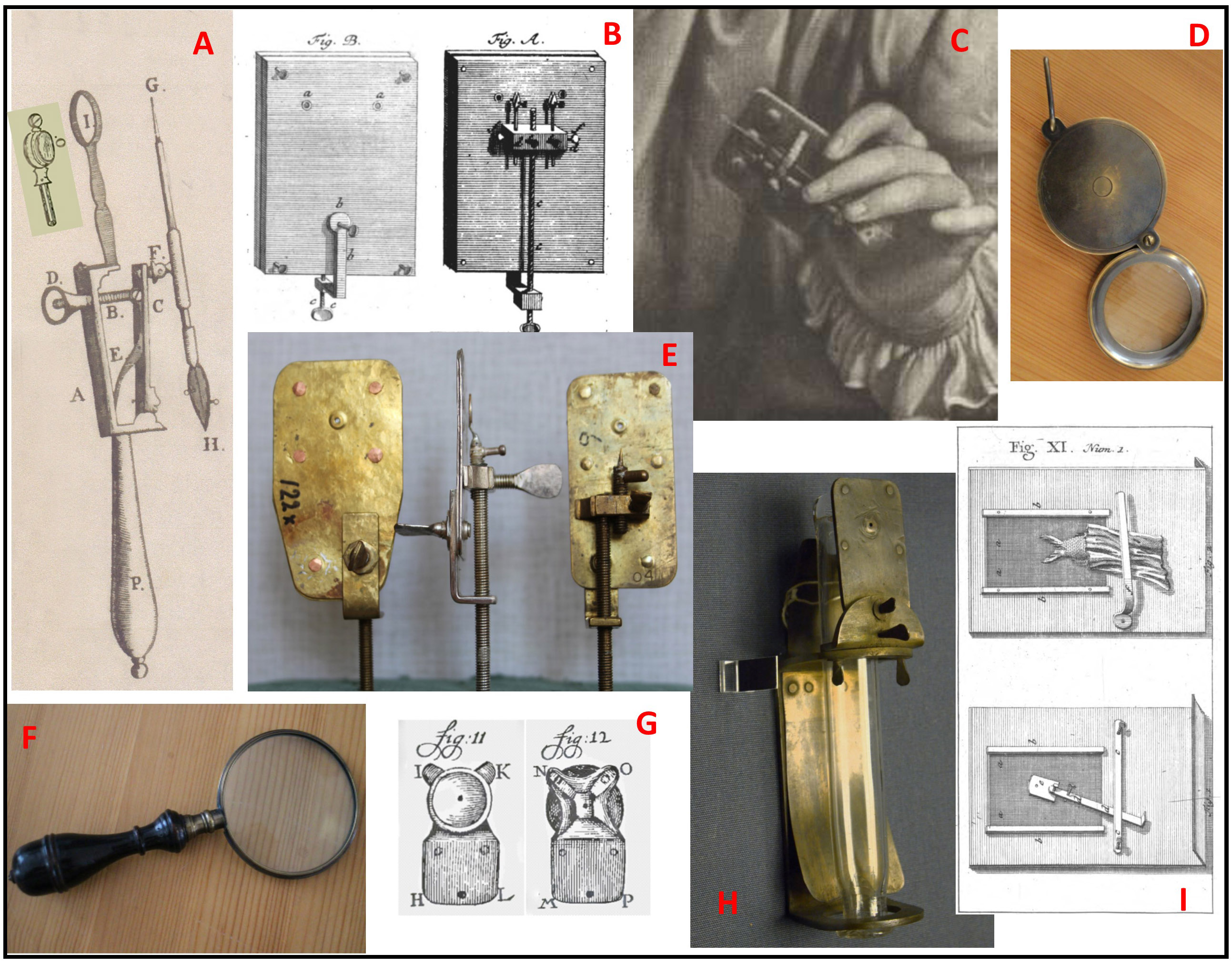
Figure 2: Optical toolbox
A: This magnifying glass (or something very like it) appears in the frontispiece of the auction catalogue for the sale of Van Leeuwenhoek’s microscopes after his daughter’s death. Because the lens is set into a simple ring rather than the metal plate of the Van Leeuwenhoek microscope, the setting does not cast a shadow on the sample if reflected lighting is used.
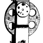
B: This version of the Van Leeuwenhoek microscope was published by a German visitor (Zacharius-Conrad von Uffenbach), and microscopes with 2 lenses are mentioned in the sale catalogue. With 2 different lenses side by side, it would have been more convenient than moving a fragile sample to another microscope for a different magnification.
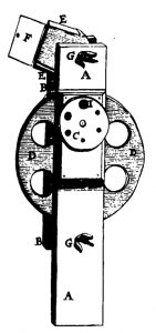
C: This 3-lensed microscope comes from an engraving made while Van Leeuwenhoek was in the prime of life, and is also mentioned in the catalogue. Note the holder for a capillary (used for liquid samples such as blood) beside the sample needle.
D and F: Copies of 17/18th century magnifying glasses, as sold by the Rijksmuseum Boerhaave. “Burning glasses” are mentioned in the inventory of the Van Leeuwenhoek house after Maria van Leeuwenhoek’s death. As well as magnifying larger samples, they can be used to control the lighting of samples on the microscopes.
E: Some of the facsimiles of existing Van Leeuwenhoek microscopes made by Hans Loncke and used in my experiments.
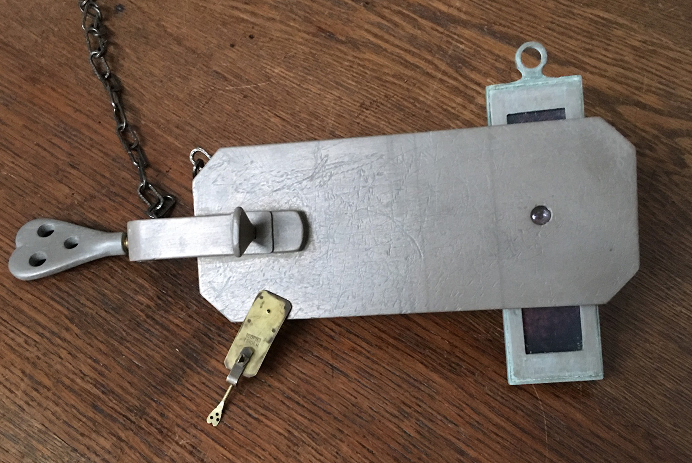
G: Van Leeuwenhoek’s adaption of the eyepiece on his aalkijker to allow reflected lighting while protecting the eye with a small cup.
H: A facsimile of the aalkijker drawn in Van Leeuwenhoek’s first paper on blood circulation. Note the use of the same lens plate as with the microscopes.
I: His final adaption of the aalkijker to make its use easier for visitors, as mentioned by him in a letter and drawn by Von Uffenbach.
People often describe his microscopes as crude, and perhaps they are when compared to other, ornamented microscopes of his time, but I suspect that Antoni van Leeuwenhoek was more interested in making something that would allow him to carry out his experiments rather than producing elegant instruments.
Exploring the Delft School’s microscope collection – 1.
As I have mentioned before, the Delft School of Microbiology Archive has a small collection of old and unusual microscopes. We also have a range of attachments with varying (and sometimes unidentified) functions as well as two boxes of mysterious parts. One of my projects for this winter is to try as many of these things out as possible, and write a methods book for the collection.
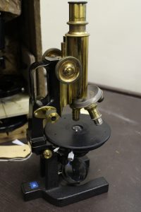
I have started with two red boxes from Ernst Abbe’s time at Carl Zeiss Jena (CZJ). Abbe was one of the founding fathers of CZJ and the inventor of many microscope techniques that we take for granted today, including standardizing lens quality. One of the boxes contains the parts necessary to convert a simple microscope for work with polarization, the other contains a camera lucida. Both were obviously routinely used in the times of Beijerinck, Van Iterson and Kluyver and we have enough of each for every student in a practical class to use them. None of the boxes included instructions, and nobody I’ve asked had tried to use either one. Since they turn up sometimes on auction sites, it seems to be worth outlining my findings here. I used my late 19th century jug-handled CZJ microscope.
-
- Abbe's polarizer
-
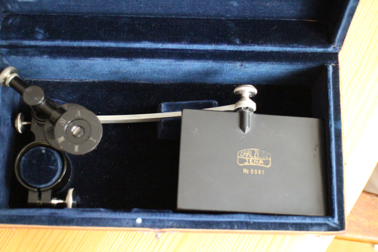
- Abbe's camera lucida
-
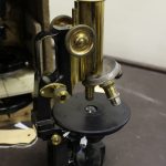
- My CZJ microscope
The polarizer.
The box contains a polarizer (top right in the photo), a calibrated ring (centre) and an eyepiece (bottom left). It took me a while to realize that the polarizer is actually two pieces which unscrew. The piece with the lens fits into the filter ring under the condenser, and the ring screws on underneath the filter ring to stabilize the polarizer. The calibrated ring fits around, and the eyepiece over the ocular.
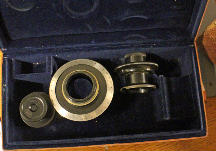
The contents of the polarizer box.
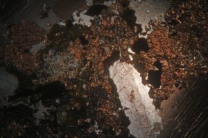
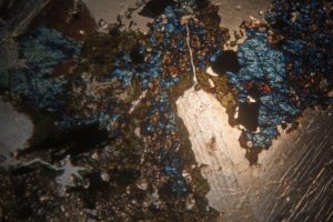
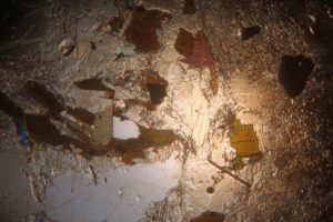
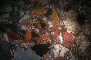
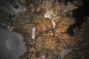
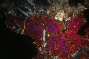
Using my usual LED lamp, I adjusted the mirror and condenser to give optimum light and then inserted a slide. Rather than microorganisms, for these experiments it was simpler to use samples guaranteed to give the dramatic colour changes associated with polarizing microscopy and so I chose mineral samples prepared for the microscope by Dr F Krantz of Bonn around the beginning of the 20th century (and also in our collection). The pairs of photos show the extremes of uncrossed and crossed polarized light paths for 3 different minerals.
The camera lucida
The box contains a ring which fits over the microscope’s ocular. Attached to the ring is an arm with a mirror at its end and a shorter, moveable arm supporting the prism that combines the images from the microscope and the mirror. The arm allows the prism to swing over or away from the ocular, allowing the microscope to be used with and without it (very useful for focusing and sample placing).
Initially, I found this attachment very frustrating because everyone I had discussed it with confidently said that it projected the sample’s image onto paper beside the microscope. This did not happen. It was only when I looked through the ocular to check the microscope’s focus and saw a ghostly pen superimposed on the sample that I realised that the projection was the other way round! Thus far, I am not very satisfied with my photographs of the combined images, but I’ll post a picture here when I’m happy with them. The problem is not in using the equipment, but in convincing the camera that it can focus on the pen and sample at the same time – I see that CZJ’s catalogues of the time also offer a drawing platform for use with their camera lucida which was presumably exactly the correct height. If anyone wants to try, I’ve had better results with cream coloured paper rather than bright white paper which tends to reflect more in the field of view. Reducing the light in the room also helps.
Van Musschenbroek Microscopes
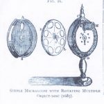
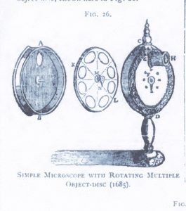
It cannot be denied that Antoni van Leeuwenhoek made major discoveries with his single lensed microscopes. Consequently, when thinking about the simple microscopes of the 16th and 17th centuries, most people have focused on his very simple (some say crude) microscopes. In fact, there were several other single lensed microscopes in use at the time. Many of them were more complicated than Van Leeuwenhoek’s microscope with extra features and even engraved ornamentation. One had a rotating disc with different sized holes to give varying apertures, others had detachable lenses or moving sample holders so that samples did not have to be transferred to another microscope for a change of magnification. Significant discoveries were also made with these microscopes and yet they are rarely studied. Comparison of results from the different types could tell us so much more, but working replicas (with proper lenses) like the Van Leeuwenhoek replicas I normally work with are not to be found.
-
- Kircher
-
- Huygens 1
-
- Huygens ver 2
-
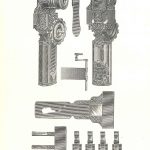
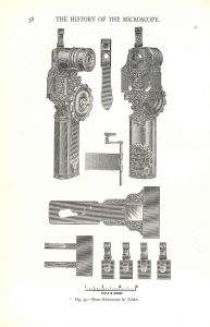
- Joblot
-
- Hartsoeker

-
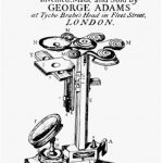
- Adams

Last year, after I complained about the lack of such replicas for experimental use during a lecture, someone called my bluff. Dr Koen Quint of the Dutch Stichting voor Historische Microscopie offered me the honour of trying their (real) Van Musschenbroek microscope! I have to admit that my first reaction was panic.
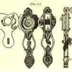
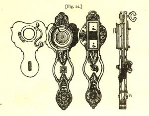
The Van Musschenbroek workshop in Leiden was a family business between 1660 and 1750. They produced (and invented) a range of scientific instruments, among which were microscopes. At its peak between 1730 and 1740, their microscope range included 10 different models, some of which were their own invention. The more complicated of their single lensed microscopes included a holder and detachable lenses.

Van Musschenbroek microscope with lenses and sample holders
The initial experiments have been encouraging. Of course, using a centuries-old microscope was a bit nerve wracking, and much of the first session was spent working out how to set everything up without damaging it (e.g. we wrapped the handle of the microscope in felt to protect it against the clamps holding it erect). Obviously I can’t take it home to use on wet Sunday afternoons as I would with a replica, but the next few weeks are going to be fun!

Experimental setup
Van Iterson’s wall chart collection.
In the days before the easy projection of images, wall charts were the usual manner of illustrating lectures and practical classes. This photo shows the autumn 1941 practical on microscopical research of living plants and plant products, with wall charts in use on the back wall. Prof van Iterson can be seen standing in the far left corner of the room.
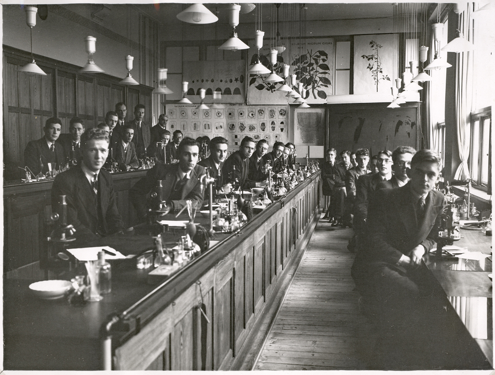
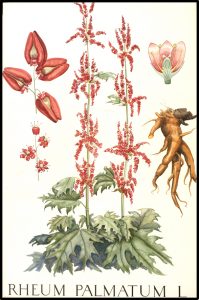
The Archive has a large collection of these wall charts, and I’ve previously shown some of the printed ones as well as some by Henriette Beijerinck. The original wall charts for Van Iterson’s Department were produced by at least 15 people, not all of whom signed their work with their full names. They range from beautiful watercolours of useful plants to diagrams and tables – everything a Professor of Applied Botany could need for his lectures.
-
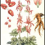
- Ornamental rhubarb by Karin Mauve, 1920s
-
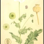
- Poppy by Hilda Kern, 1940s or 50s

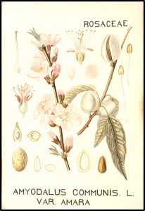
-
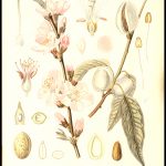
- Almonds by ? Bongers, 1923
-
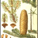
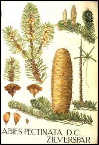
- Silver spar pine by J. Mouton, 1920s
-
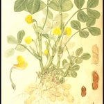
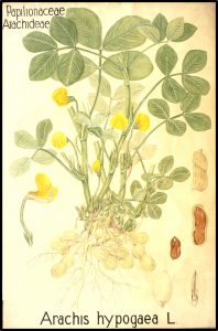
- Peanuts by Sluyterman, 1920s
-
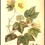
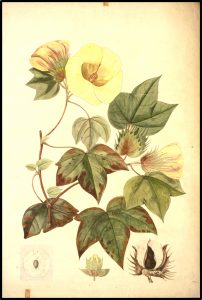
- Cotton by Karin Mauve, 1920s
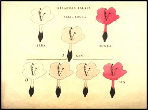
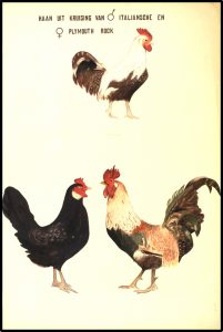
Like Beijerinck and Kluyver, in those days before the discovery of DNA, Van Iterson was interested in heredity and variation, and we have a series of watercolours illustrating Mendelian genetics, gender dimorphism and mimicry.
-
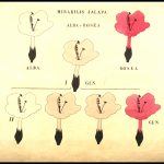
- Mendelian cross of white and pink Mirabilis jalapa. Unknown.
-
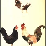
- Cross of Italian and Plymouth Rock chickens
-
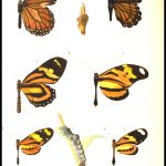
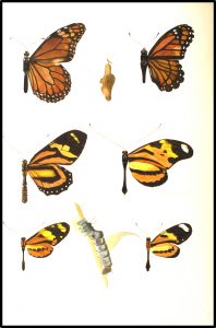
- Butterfly mimicry by J. Mouton, 1920s?
Researching and cataloguing the wall chart collection was a great labour of love by Truus ten Hoopen-van Hulsentop. If anyone can tell us anything more about the various artists, we’d be delighted to hear from you.
Something Van Leeuwenhoek didn’t see!
On 7th May (2017) the Dutch Society for Microscopy (Nederlands Genootschap voor Microscopie) ran an open microscopy workshop at the Delft Science Centre. They taught members of the public (and me!) how to make thin sections of plant material, stain the sections and then examine them under modern microscopes.
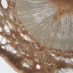
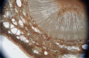
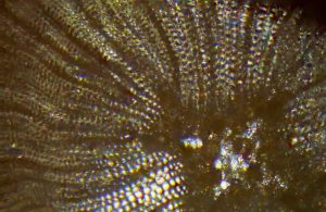
The plant material in question was a twig from Wollemia nobilis (captive bred). W. nobilis is a very rare pine tree that was only known from the fossil record until 1994 when a few trees were found in Australia. The oldest tree has been estimated to be over 1000 years old according to the Kew Gardens website. As the only representatives of a genus that dates back to the time of the dinosaurs, the wild trees are IUCN-red listed and stringently protected, but seedlings can now sometimes be bought.
Since there were a few sections left at the end of the workshop, how could I resist putting them under my 65x facsimile Van Leeuwenhoek microscope? He would, of course, have examined them if they had been available to him 300 years ago.
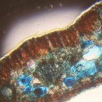
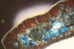
A couple of images using a much weaker lens on my 19th century Zeiss “jug handled” microscope have also been included for comparison (the first photo on each line). The red and blue images came from a stained preparation, the others from a section that had been allowed to dry on a coverslip, without staining.
-
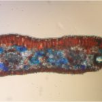
- Stained needle section under the Zeiss
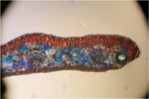
-
- Stained needle section under the AvL
-
- Stem section under the Zeiss
-
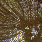
- Central area of stem section under the AvL
-
- Outer area of stem section under AvL

Antoni van Leeuwenhoek in Spain
I have just heard that it will be possible to see part of the beautiful Camacho & Pallas microscope collection at the Museum of Evolution in Burgos (Castille & Leon) in Spain.
http://www.museoevolucionhumana.com/
The exhibition will run from April to November, 2017.
This is a privately-owned collection which includes the Van Leeuwenhoek microscope that was found in mud dredged from Delft’s Oude Delft canal in in 2015 (see my blogpost from 23 June 2015).
If you’re going to be holidaying in Spain this summer, it’ll be well worth a visit.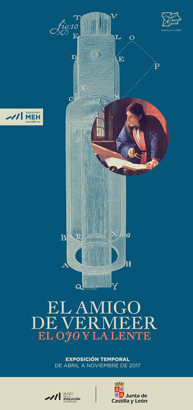
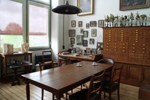
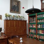
Beijerinck’s office (and the neighbours)…
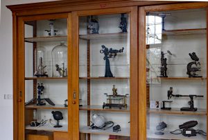
At long last, the Beijerinck Museum is ready for visitors – indeed several groups, including the Dutch Microscopy Society and guests from the Queckett Microscopical Club, have had sneak previews. Also known as “Beijerinck’s Office” (Kamer van Beijerinck), it will only be open to escorted groups as we don’t want to spoil the atmosphere by putting everything into locked display cabinets. Some of our microscopes are now on display in the foyer of the Mekelzaal, the main conference room of the Science Centre.
-
- Beijerinck's office 2
-
- Beijerinck's office 1

-
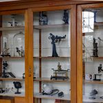
- Microscope display
-

- Monuments to Beijerinck, Van Iterson, Kluyver and Van Rossem
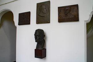
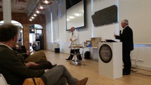
At the end of BioDay, a meeting organised to celebrate the TUDelft’s 175th birthday, the Museum was re-opened by Professor Karel Luyben, Rector Magnificus of the University, ably (?) assisted by someone claiming to be Prof Beijerinck’s assistant…
-
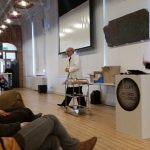
- Opening the museum
-

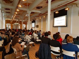
- Bioday
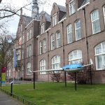
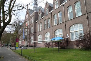
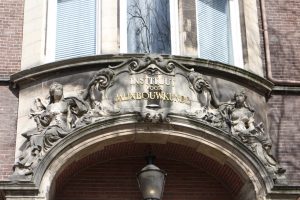
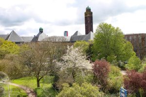
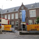
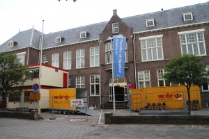
This seems like a good moment to take a look at our new surroundings. We are housed on the second floor of the Delft Science Centre. The building was originally the Faculty of Mining. It’s very easy to feel at home here as, like Prof van Iterson’s section of our previous building, our new home dates from the early 20th century and the design is very similar. The view from our windows is a great improvement!
-
- Science centre
-
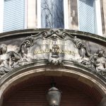
- Mining
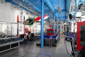
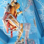
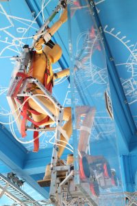
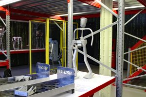
The main part of the Science Centre displays the modern achievements of Delft University of Technology and a range of prototypes (including Delft’s famous solar powered car) can be seen as well as displays that can be operated by the visitors. You can try improving the shape of an aircraft wing or use the driving simulators, among many other things – the exhibition often changes. The robotics lab is ever popular, as are the workshops where school groups (among others) come to try things out. For example, the Dutch Microscopy Society will be running a public workshop (about how to make botanic preparations (Wollemia nobilis) and look at them under the microscope) from 14:00-16:30 on 7th May (2017). Participants will only pay the Science Centre entrance price. The details are here: www.sciencecentre.tudelft.nl/nl/bezoek/agenda/event/detail/gratis-microscopie-workshop/. It’s in Dutch but the site will allow you to copy the text for pasting into Google translate (which is always amusing….).
-
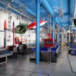
- The Science Centre's main hall
-
- climber
-
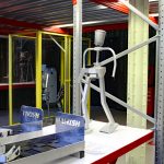
- Robotics
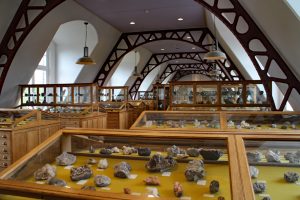
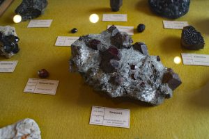
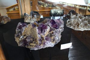
The TUDelft has always had a number of internationally important collections which started life as the working tools and products of its research departments. Among them, the minerals collection from the Faculty of Mining (which dates from the middle of the 19th century) is remarkably beautiful. It is housed, in its original display cases, in rooms next door to the Beijerinck Museum and in cabinets in other public parts of the building. Like the Beijerinck Museum, the Minerals Collection is open to escorted groups.
-
- Minerals museum
-
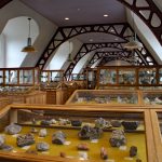
- Minerals exhibition
-
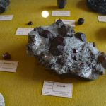
- Garnets
-
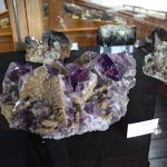
- Amethyst
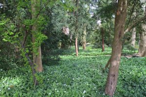
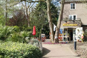
Just around the corner from the Science Centre is Delft’s Botanic Garden. Founded by Prof van Iterson with support from Prof Beijerinck, it was set up to provide plants for research in the Department of Applied Botany and is 100 years old this year (2017). Apart from their permanent collection (which includes an apiary), they frequently have exhibitions ranging from pottery through products made from plant material to photography. Their website is here: http://www.botanischetuin.tudelft.nl/en/
-
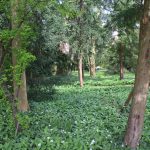
- Botanic garden
-
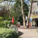
- Garden entrance
The Science Centre’s website with opening times and other information is here http://www.sciencecentre.tudelft.nl/en/.
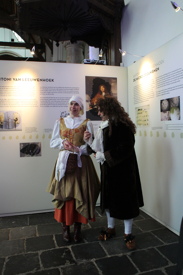
Delft, the Home of Microbiology
An exhibition at Van Leeuwenhoek’s resting place.
2017 is the 175th anniversary of the founding of the Polytechnic School that eventually became Delft University of Technology. The University is celebrating with a 175 day Lustrum, much of which will focus on the Life Sciences. It is also 100 years since Van Iterson founded the Delft Botanic Garden on the land behind his laboratory.

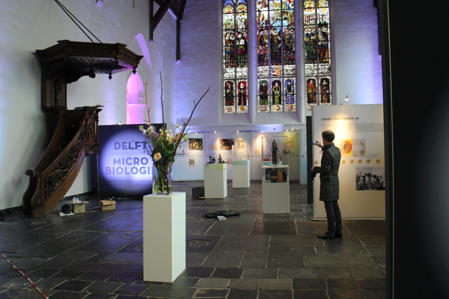
We are beginning the celebrations with a look at the history of microbiology and the biosciences in Delft, from the 17th to the 21st centuries. The exhibition will run until 26 February, 2017.

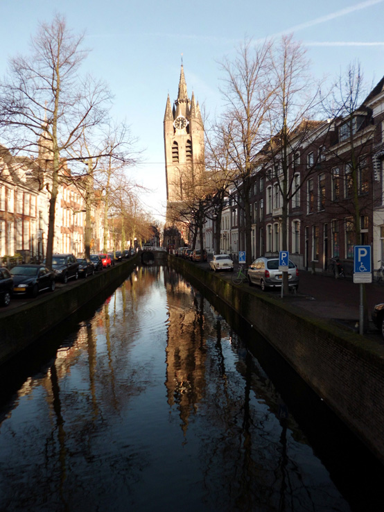
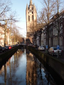
This will take the form of an exhibition in the area around Antoni van Leeuwenhoek’s grave in the Oude Kirk.
-
- Delft's Old Church
-
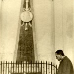
- AJ Kluyver at the grave of Antoni van Leeuwenhoek

The text on the poster boards is in Dutch, but there are also English language handouts. It includes 20th century teaching microscopes, a cross section of an electron microscope and 3-D scanned replicates of a Van Leeuwenhoek microscope and the Delft telescope.
Facsimile Van Leeuwenhoek microscope and its 3-D scanned replicate
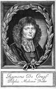
The exhibition offers a taster of the achievements of Delft microbiologists and introduces some of the people who helped and supported them. For example, we might never have heard of Antoni van Leeuwenhoek and his little animals without Reinier de Graaf, the doctor who introduced van Leeuwenhoek to the Royal Society of London, publishers of much of his work. Jacques van Marken not only brought Beijerinck to Delft to establish his industrial microbiology laboratory, he was also one of the people instrumental in creating the Department of Microbiology and Professor’s Chair for Beijerinck at what was then the Delft Polytechnic.
-
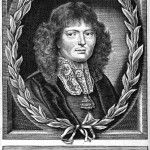
- Reinier de Graaf
Medicinae Doctor
-
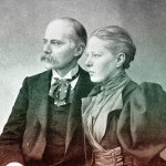
- Jan and Agneta van Marken

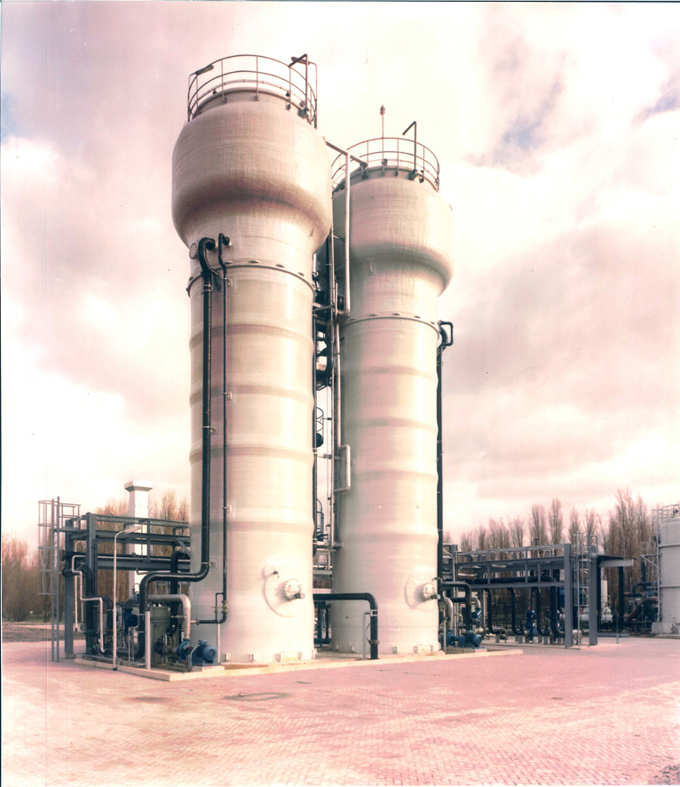
Many others have provided help, support and encouragement, but the silent contributor to the history of microbiology is the City of Delft itself. Many very well-studied microorganisms were found for the first time in samples from Delft’s canals, soil and from industrial sources, even now.
-
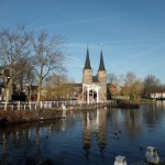
- Th old city gate
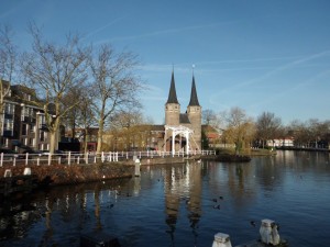
-
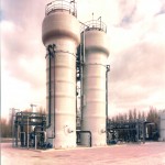
- Wastewater bioreactors

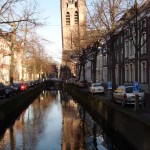
Over 300 years ago, before Van Leeuwenhoek found his little animals, the city was already a hotspot in scientific research-for example Stevin and de Groot dropped different lead balls off the tower of the New Church and proved that they hit the ground at the same time 3 years before Galileo did the same experiment from the tower of Pisa!
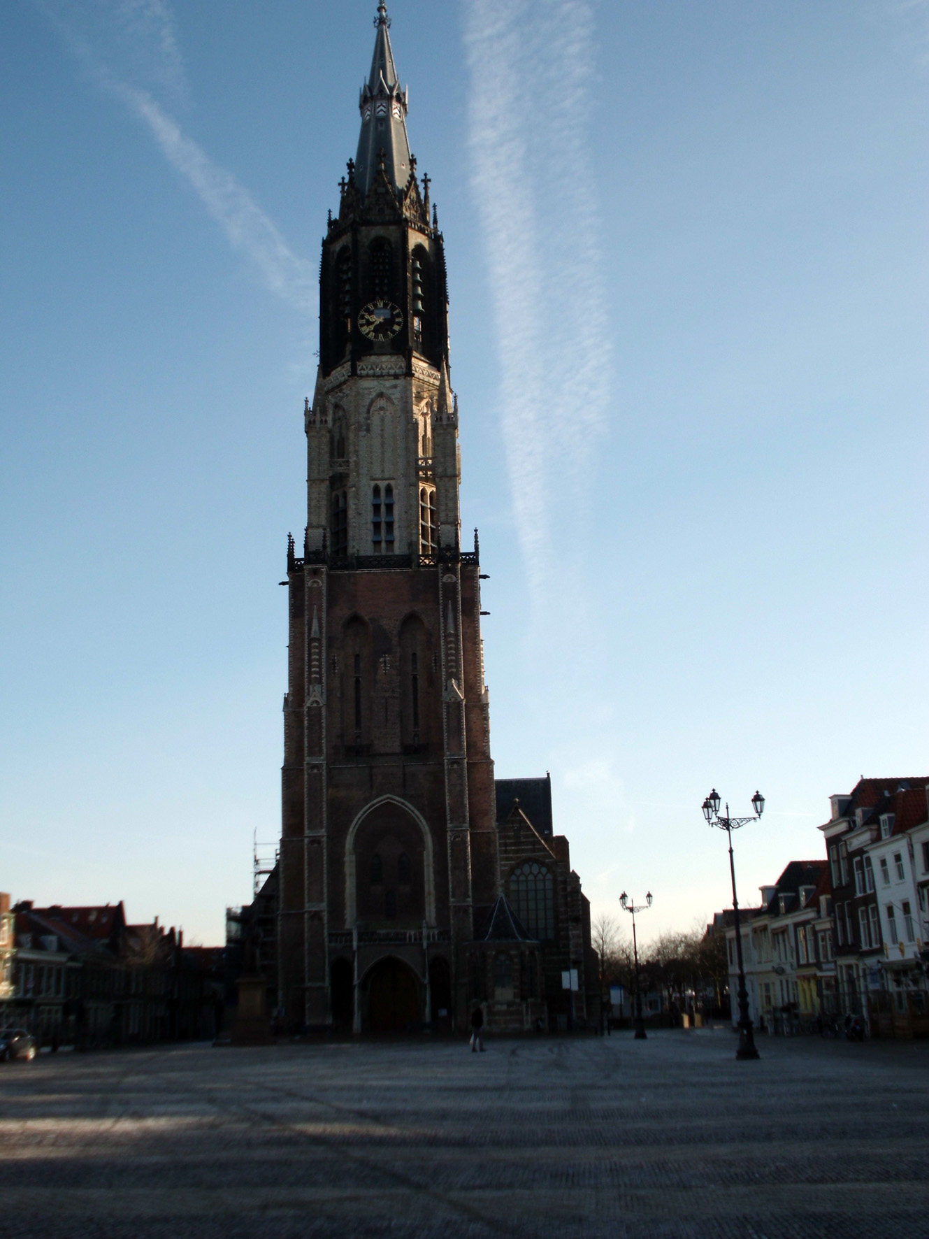
Delft’s New Church
Auld lang syne, new beginnings and a mystery solved
After weeks of chaos, mountains of used packing paper and bubble wrap, disappearing essentials and wandering packing boxes, we’re beginning to see the end of the chaos… The regular visits to the old lab to look for misplaced odds and ends have finished, and the building is now occupied by asbestos removal experts. Curiously, the information monitors are still announcing lectures and other happenings!
-
- The asbestos removers have moved in.
-
- But the monitors carry on!
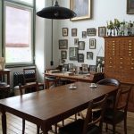
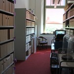
Meanwhile, back at the Science Centre, order has (mostly) appeared from chaos. Small jobs such as picture hanging still need to be done, but we can actually find things and we’ve already had our first overseas visitor!
-
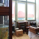
- Room with a view!

-
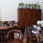
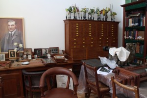
- Photos of Kluyver's contacts waiting to be hung
-
- Slowly getting sorted out

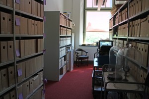
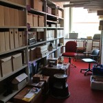
As you can see from the photos, the museum and archive spaces are a little smaller, but we have a good cellar for safe storage of items that we don’t need to use very often, so things don’t look as cluttered as they did. Working in well-lit rooms doesn’t feel quite natural yet – those who visited the old museum will remember that we had tiny windows covered with thick green anti-UV blinds and permanent electric lighting. Thanks to UV-resisting film on the glass, we can now see out of the windows, and even work by daylight!
-
- Equipment waiting for just the right place..
-
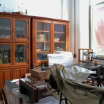
- Archive boxes and the wall chart cabinets

-
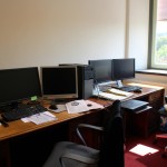
- The luxury of an office
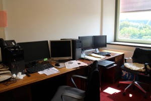
We even have a proper office instead of odd desks squeezed in wherever we could – luxury!
Mystery solved
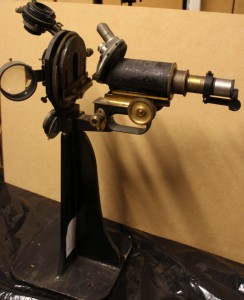
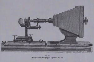
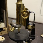
Returning to the strange microscope of my previous post, I think that I have solved the mystery. While cleaning it, I noticed that the heavy stand is not of the same quality as the rest of the microscope, and (unlike the rest of the microscope parts) does not have the Carl Zeiss Jena (CZJ) name or number. Also, the screws and hooks holding the four lenses and condensers are a bit rusty (and thus not up to CZJ’s standard). I then came across the diagram below in the 1885 CZJ catalogue, and realised that what we have is a home-made version. The microscope in the diagram only has a single condenser, but whoever made ours obviously felt that if a thing was worth doing, it was worth overdoing!
-
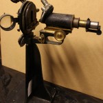
- Our home made version
-
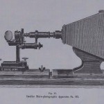
- Photo microscope from CZJ in 1885
-
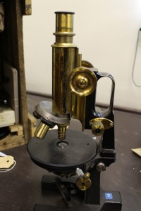
- 19th century "jug handled" Zeiss microscope
-
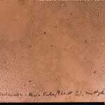
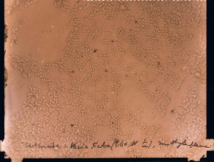
- 1880s Glass negative showing Rhizobium
What we have is a standard CZJ “jug handled” microscope from the end of the 19th century, lying on its back and converted for micro-photography. Sadly, we don’t have the camera, but it does explain how the early photographs (beginning in the 1880s) in our collection of glass negatives were made.
Recent Comments