
Posts in category Uncategorized
Something Van Leeuwenhoek didn’t see!
On 7th May (2017) the Dutch Society for Microscopy (Nederlands Genootschap voor Microscopie) ran an open microscopy workshop at the Delft Science Centre. They taught members of the public (and me!) how to make thin sections of plant material, stain the sections and then examine them under modern microscopes.
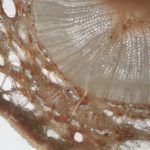
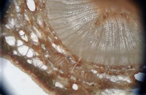
The plant material in question was a twig from Wollemia nobilis (captive bred). W. nobilis is a very rare pine tree that was only known from the fossil record until 1994 when a few trees were found in Australia. The oldest tree has been estimated to be over 1000 years old according to the Kew Gardens website. As the only representatives of a genus that dates back to the time of the dinosaurs, the wild trees are IUCN-red listed and stringently protected, but seedlings can now sometimes be bought.
Since there were a few sections left at the end of the workshop, how could I resist putting them under my 65x facsimile Van Leeuwenhoek microscope? He would, of course, have examined them if they had been available to him 300 years ago.
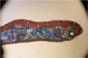
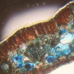
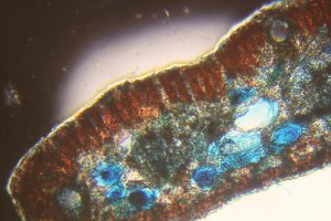
A couple of images using a much weaker lens on my 19th century Zeiss “jug handled” microscope have also been included for comparison (the first photo on each line). The red and blue images came from a stained preparation, the others from a section that had been allowed to dry on a coverslip, without staining.
-
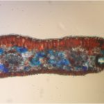
- Stained needle section under the Zeiss
-
- Stained needle section under the AvL
-
- Stem section under the Zeiss
-
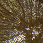
- Central area of stem section under the AvL
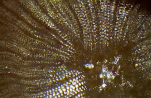
-
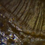
- Outer area of stem section under AvL

Antoni van Leeuwenhoek in Spain
I have just heard that it will be possible to see part of the beautiful Camacho & Pallas microscope collection at the Museum of Evolution in Burgos (Castille & Leon) in Spain.
http://www.museoevolucionhumana.com/
The exhibition will run from April to November, 2017.
This is a privately-owned collection which includes the Van Leeuwenhoek microscope that was found in mud dredged from Delft’s Oude Delft canal in in 2015 (see my blogpost from 23 June 2015).
If you’re going to be holidaying in Spain this summer, it’ll be well worth a visit.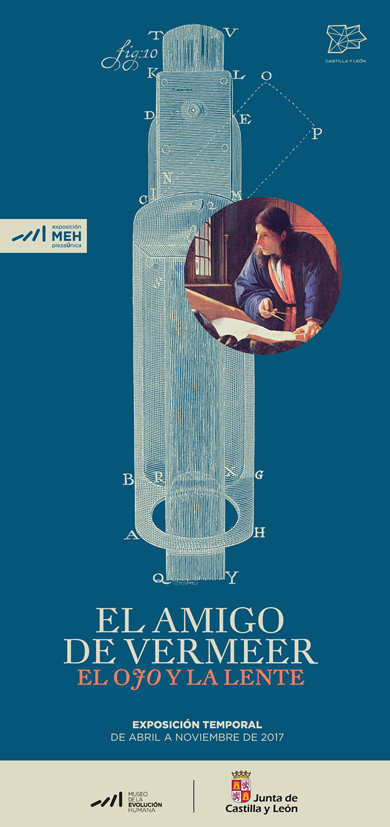
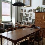
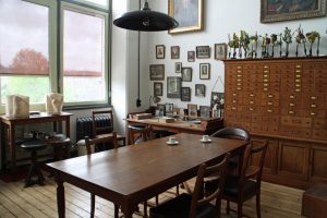
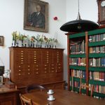
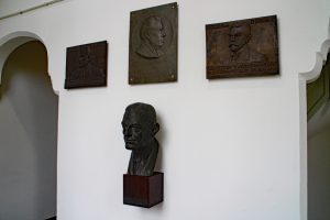
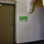
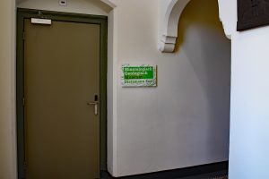
Beijerinck’s office (and the neighbours)…
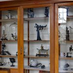
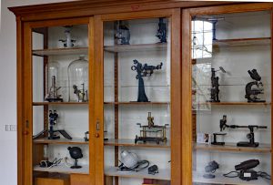
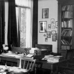
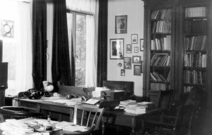
At long last, the Beijerinck Museum is ready for visitors – indeed several groups, including the Dutch Microscopy Society and guests from the Queckett Microscopical Club, have had sneak previews. Also known as “Beijerinck’s Office” (Kamer van Beijerinck), it will only be open to escorted groups as we don’t want to spoil the atmosphere by putting everything into locked display cabinets. Some of our microscopes are now on display in the foyer of the Mekelzaal, the main conference room of the Science Centre.
-
- Beijerinck's office 2
-
- Beijerinck's office 1

-
- Microscope display
-

- Monuments to Beijerinck, Van Iterson, Kluyver and Van Rossem
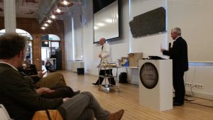
At the end of BioDay, a meeting organised to celebrate the TUDelft’s 175th birthday, the Museum was re-opened by Professor Karel Luyben, Rector Magnificus of the University, ably (?) assisted by someone claiming to be Prof Beijerinck’s assistant…
-
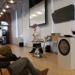
- Opening the museum
-
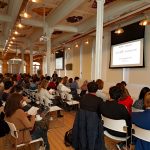
- Bioday
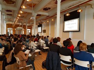
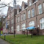
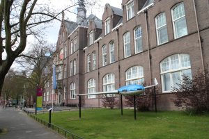
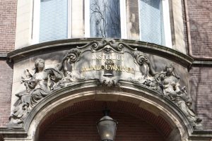
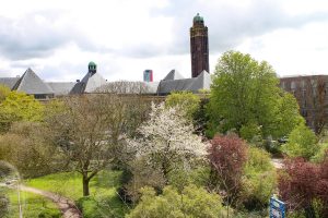
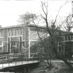
This seems like a good moment to take a look at our new surroundings. We are housed on the second floor of the Delft Science Centre. The building was originally the Faculty of Mining. It’s very easy to feel at home here as, like Prof van Iterson’s section of our previous building, our new home dates from the early 20th century and the design is very similar. The view from our windows is a great improvement!
-
- Science centre
-
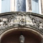
- Mining
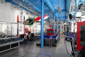
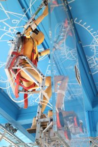
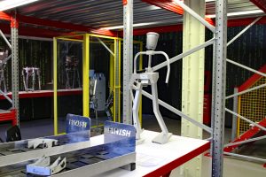
The main part of the Science Centre displays the modern achievements of Delft University of Technology and a range of prototypes (including Delft’s famous solar powered car) can be seen as well as displays that can be operated by the visitors. You can try improving the shape of an aircraft wing or use the driving simulators, among many other things – the exhibition often changes. The robotics lab is ever popular, as are the workshops where school groups (among others) come to try things out. For example, the Dutch Microscopy Society will be running a public workshop (about how to make botanic preparations (Wollemia nobilis) and look at them under the microscope) from 14:00-16:30 on 7th May (2017). Participants will only pay the Science Centre entrance price. The details are here: www.sciencecentre.tudelft.nl/nl/bezoek/agenda/event/detail/gratis-microscopie-workshop/. It’s in Dutch but the site will allow you to copy the text for pasting into Google translate (which is always amusing….).
-
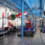
- The Science Centre's main hall
-
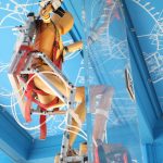
- climber
-
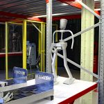
- Robotics
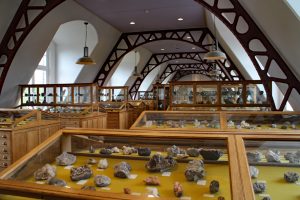
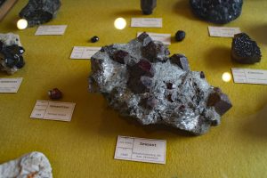
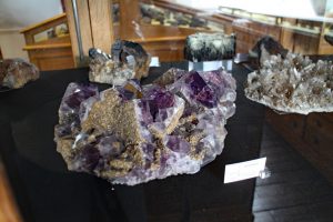
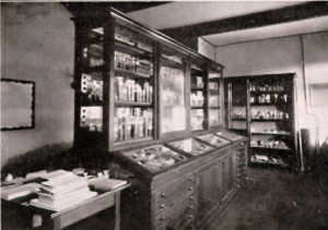
The TUDelft has always had a number of internationally important collections which started life as the working tools and products of its research departments. Among them, the minerals collection from the Faculty of Mining (which dates from the middle of the 19th century) is remarkably beautiful. It is housed, in its original display cases, in rooms next door to the Beijerinck Museum and in cabinets in other public parts of the building. Like the Beijerinck Museum, the Minerals Collection is open to escorted groups.
-
- Minerals museum
-
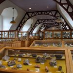
- Minerals exhibition
-
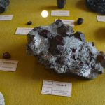
- Garnets
-
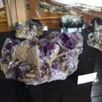
- Amethyst
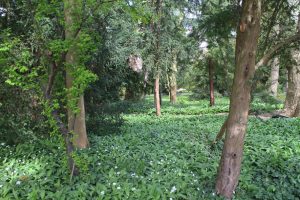
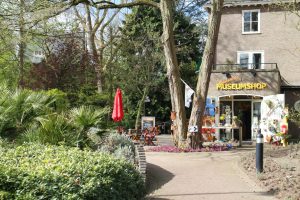
Just around the corner from the Science Centre is Delft’s Botanic Garden. Founded by Prof van Iterson with support from Prof Beijerinck, it was set up to provide plants for research in the Department of Applied Botany and is 100 years old this year (2017). Apart from their permanent collection (which includes an apiary), they frequently have exhibitions ranging from pottery through products made from plant material to photography. Their website is here: http://www.botanischetuin.tudelft.nl/en/
-
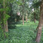
- Botanic garden
-
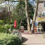
- Garden entrance
The Science Centre’s website with opening times and other information is here http://www.sciencecentre.tudelft.nl/en/.
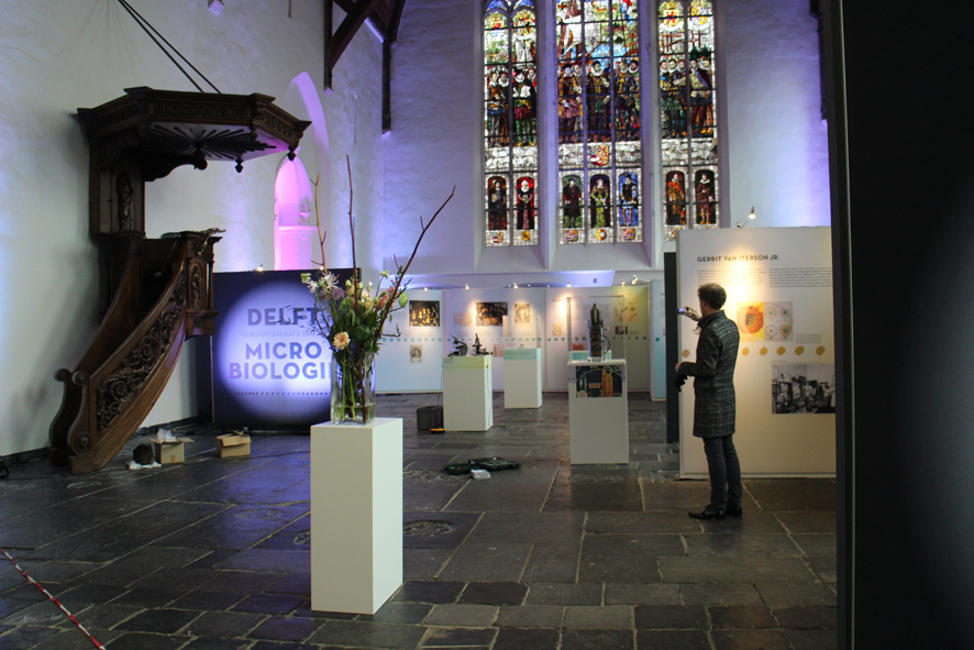
Delft, the Home of Microbiology
An exhibition at Van Leeuwenhoek’s resting place.
2017 is the 175th anniversary of the founding of the Polytechnic School that eventually became Delft University of Technology. The University is celebrating with a 175 day Lustrum, much of which will focus on the Life Sciences. It is also 100 years since Van Iterson founded the Delft Botanic Garden on the land behind his laboratory.

We are beginning the celebrations with a look at the history of microbiology and the biosciences in Delft, from the 17th to the 21st centuries. The exhibition will run until 26 February, 2017.

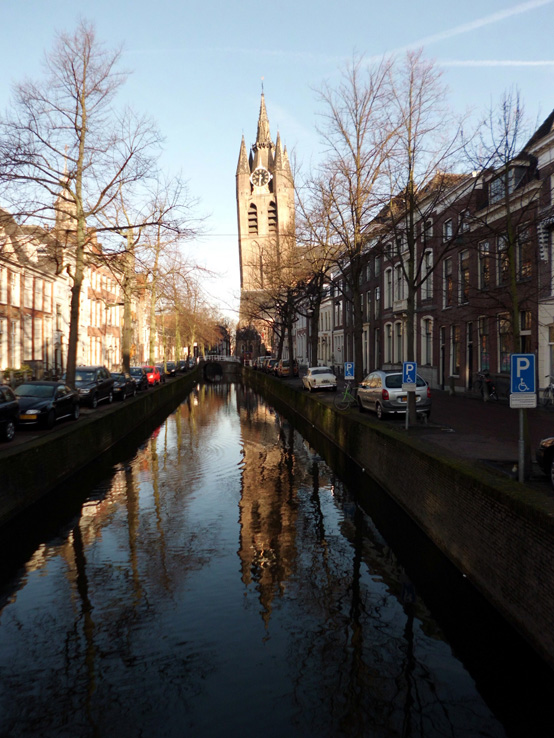
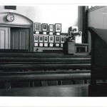
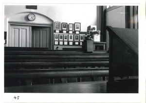
This will take the form of an exhibition in the area around Antoni van Leeuwenhoek’s grave in the Oude Kirk.
-
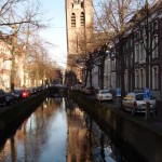
- Delft's Old Church
-
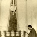
- AJ Kluyver at the grave of Antoni van Leeuwenhoek

The text on the poster boards is in Dutch, but there are also English language handouts. It includes 20th century teaching microscopes, a cross section of an electron microscope and 3-D scanned replicates of a Van Leeuwenhoek microscope and the Delft telescope.
Facsimile Van Leeuwenhoek microscope and its 3-D scanned replicate
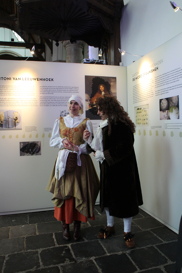
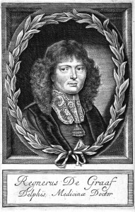
The exhibition offers a taster of the achievements of Delft microbiologists and introduces some of the people who helped and supported them. For example, we might never have heard of Antoni van Leeuwenhoek and his little animals without Reinier de Graaf, the doctor who introduced van Leeuwenhoek to the Royal Society of London, publishers of much of his work. Jacques van Marken not only brought Beijerinck to Delft to establish his industrial microbiology laboratory, he was also one of the people instrumental in creating the Department of Microbiology and Professor’s Chair for Beijerinck at what was then the Delft Polytechnic.
-
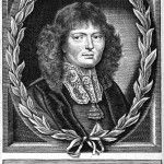
- Reinier de Graaf
Medicinae Doctor
-
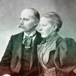
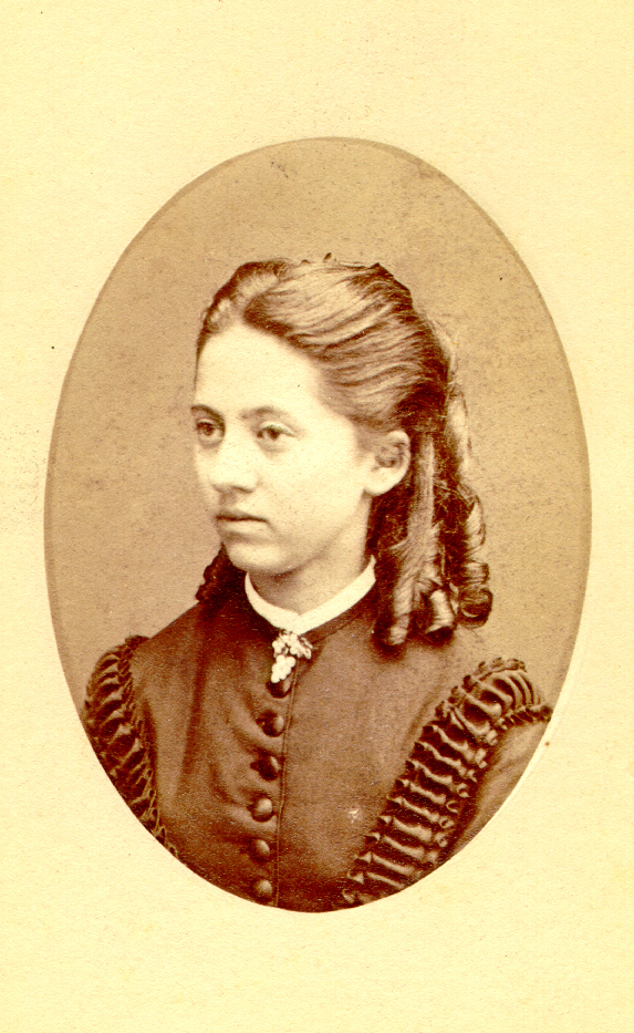
- Jan and Agneta van Marken

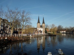
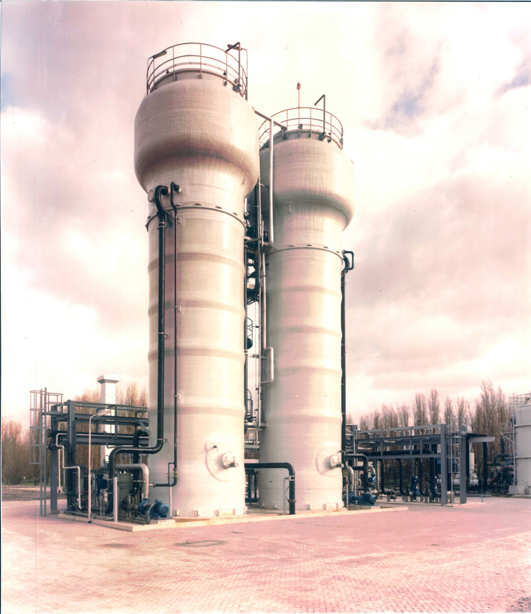
Many others have provided help, support and encouragement, but the silent contributor to the history of microbiology is the City of Delft itself. Many very well-studied microorganisms were found for the first time in samples from Delft’s canals, soil and from industrial sources, even now.
-
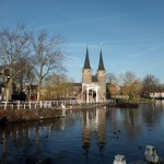
- Th old city gate
-
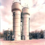
- Wastewater bioreactors

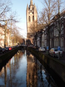
Over 300 years ago, before Van Leeuwenhoek found his little animals, the city was already a hotspot in scientific research-for example Stevin and de Groot dropped different lead balls off the tower of the New Church and proved that they hit the ground at the same time 3 years before Galileo did the same experiment from the tower of Pisa!
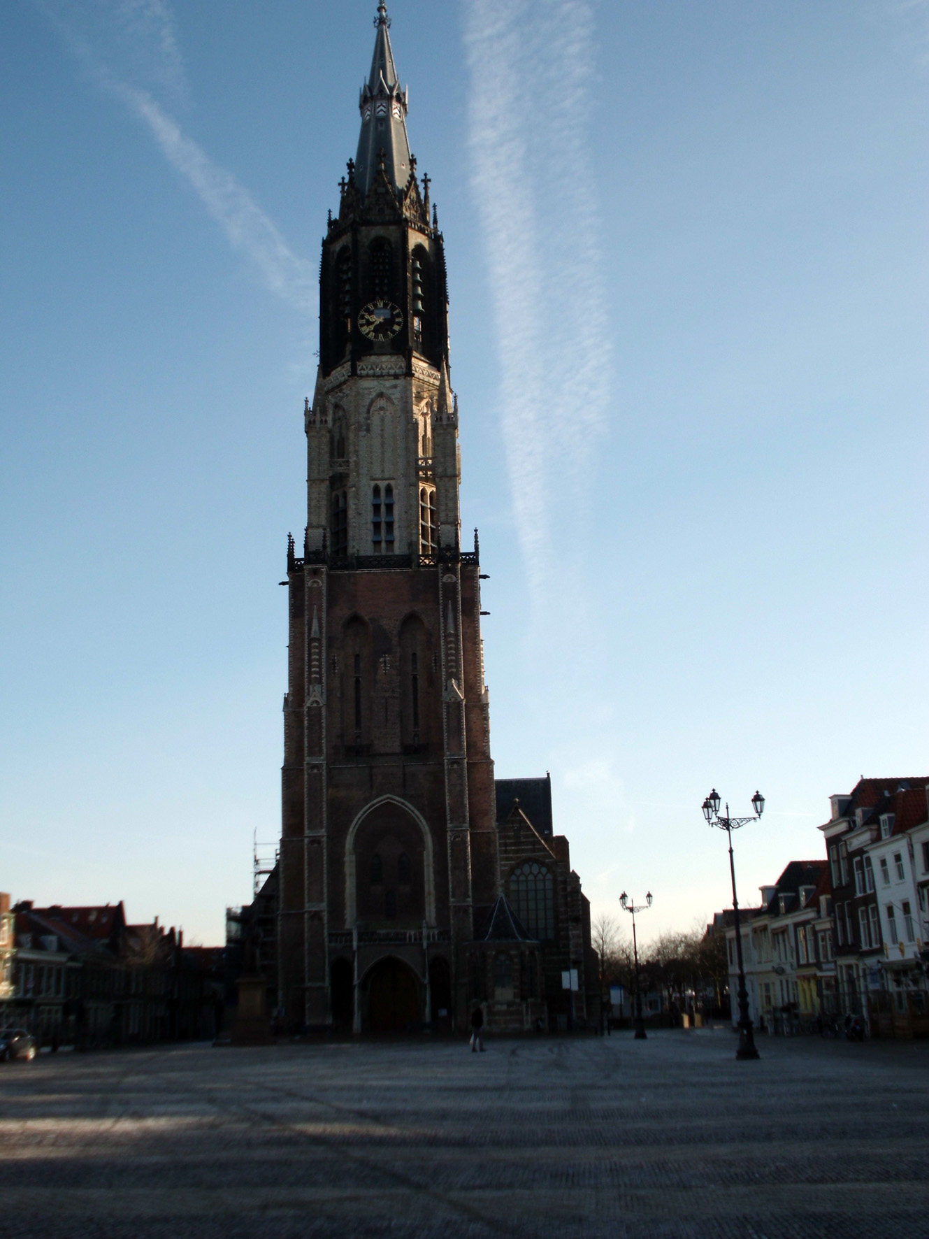
Delft’s New Church
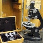
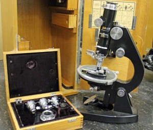
What was this microscope used for?
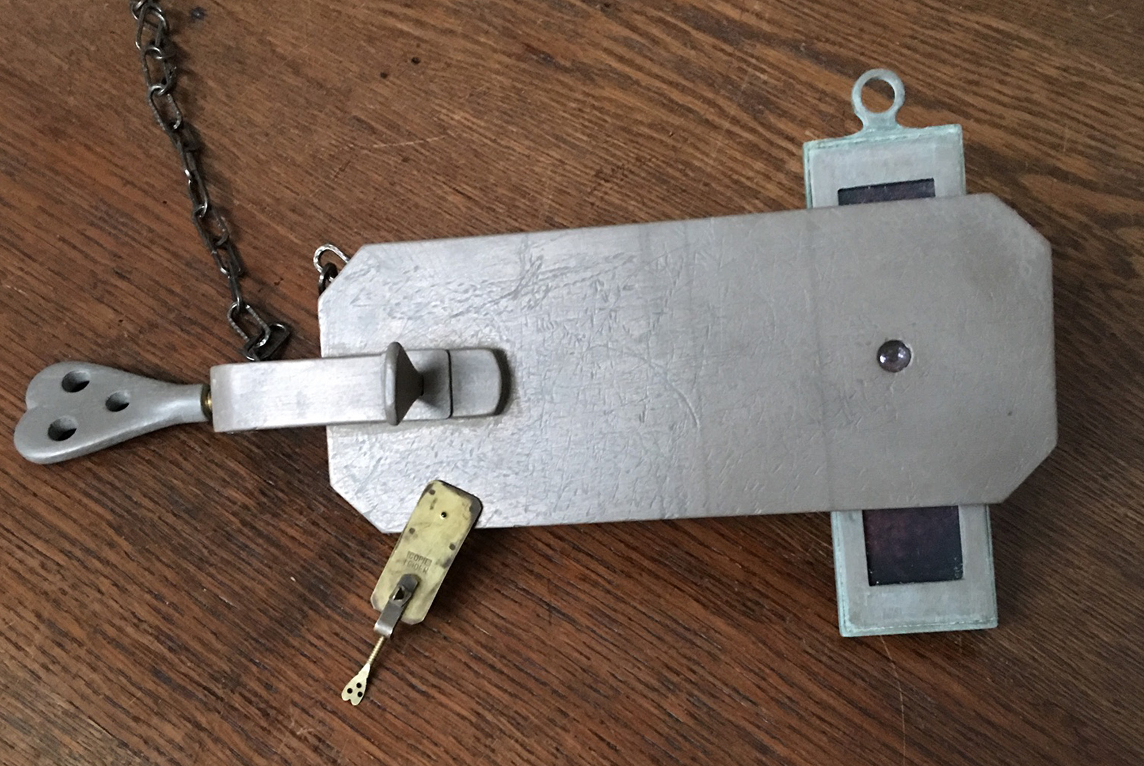
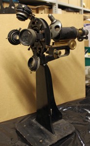
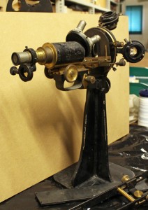
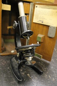
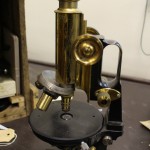
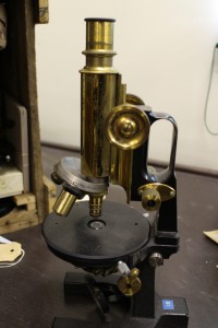
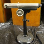
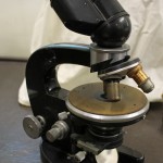
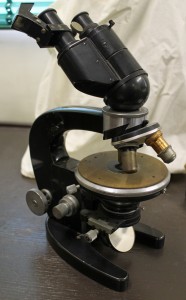
Discovering rare or unusual microscopes has become almost routine since we began packing the collection, but this one has me (and everyone else who has visited) baffled.
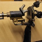
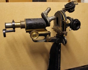
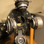
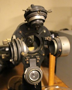
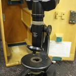
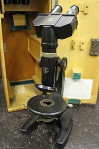
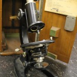
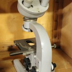
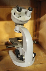
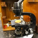
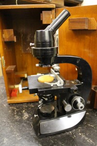
It seems to be a “jug handled” Carl Zeiss Jena microscope from the late 19th/early 20th century. However, it is mounted on its back rather than on a foot (1). Moreover, in place of the usual single condenser, there’s what seems to be 2 lenses of different strengths and 2 condensers (2), hinged so that any of them can be used to light the preparation (3). There’s an additional lens which can swing over the usual ocular (4).
Has anyone any idea what it was used for?
-
- 1: side view
-
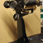
- 2: "foot"end
-
- 3: "condensers"
-
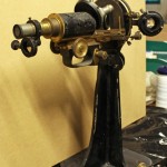
- 4: ocular end
From “out of date junk” to “exciting and rare” – our microscope collection
At the moment, sorting out the cupboards before our move has become very exciting as I’ve reached the microscope collection. Much of it was stored in the 1950s when the Laboratory of Microbiology moved from its original building, and has rarely been disturbed since then. Some of the microscopes date from the late 19th century, and even some of the 20th century ones are more interesting than might be expected.
The youngest of the companies represented in our microscope collection is probably the least well-known, especially outside the Netherlands.
Bleeker Nedoptifa
Founded by Dr Caroline E. (Lili) Bleeker and Gerard Willemse in 1939, Nedoptifa rapidly became known for the high standard of their optical products. They began with the production of binoculars for the Dutch army, but production was interrupted by WW2. Most of their microscope production seems to have been after 1945, when the “Nedoptifa” name came into use. The company cooperated with the Nobel prize-winner, Frits Zernike, in the development of phase contrast microscopy and held his patent on phase contrast microscopes. Bleeker retired at the end of 1963, the company was eventually taken over by a Delft firm and then in 1978 the factory in Zeist was closed.
Kluyver’s group seems to have used the basic Nedoptifa microscope for teaching – we have quite a few of them. Most of them have the standard circular stage, but a few are square. We also have one of their very early binocular microscopes as well as monocular and binocular phase contrast microscopes. Most of them, with the exception of the binocular phase contrast microscope, seem to have been use in in the laboratory before 1955.
Note added later: Disappointingly, after a visit in mid-June from Peter Paul de Bruyn, an expert on Bleeker microscopes, it seems that the microscopes whose boxes proclaimed them to be phase contrast microscopes do not have the necessary lenses or fittings. They may appear as we complete the packing of the collection, but it’s beginning to look unlikely.
-
- Bleeker Nedoptifa binocular microscope
-
- Early Bleeker Nedoptifa "phase contrast" microscope
-
- Bleeker "phase contrast" microscope from the 1960s
Carl Zeiss Jena
This famous old microscope manufacturer needs no introduction. In what is probably our most famous portrait of Prof Beijerinck, he is clearly using one of their “jug handled” microscopes. Others in our collection are in the Zeiss catalogues of the late 19th century.
-
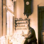
- Prof M.W. Beijerinck in his laboratory

-
- 19th century "jug handled" Zeiss microscope
We also have some more unusual examples of their work including a “horizontal microscope” (intended for examining living plants) and a binocular microscope fitted with a prism on the right ocular to aid the drawing of samples.
-
- Horizontal Zeiss microscope

-
- Zeiss drawing microscope
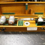
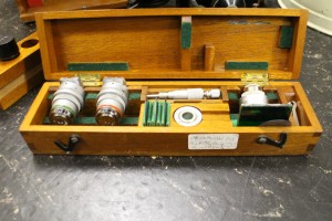
I really shouldn’t have favourites, but I must admit a fondness for the very heavy preparation microscope that turned up in a battered old wooden box on top of a cupboard. When it is taken out of the box, the eyepieces on the right flip up, giving a forerunner of our modern stereomicroscopes which can be found in the 1902 Carl Zeiss Catalogue. There are 2 sets of lenses and oculars. During dissections, the microscope can be moved backwards and forwards along the metal bar.
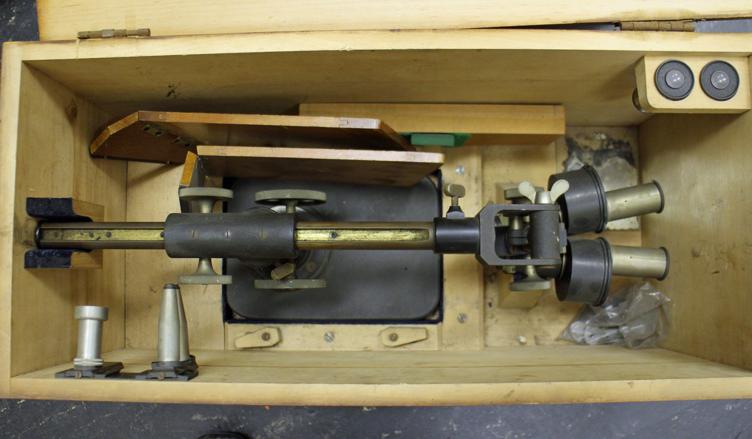
Early Zeiss preparation or dissecting microscope
Ernst Leitz Wetzlar
Another famous old company, Leitz seems to have been a favourite with Professor van Iterson. Among the microscopes from this company are a “measuring microscope”, a preparation microscope with wrist supports and the microscope (with an extensive collection of extra attachments) that he used after he retired.
-
- Leitz "measuring microscope"
-

- leitz preparation or stereo microscope

-
- Leitz microscope with one of two boxes of attachments
Other companies
Other microscope makers are mostly represented by single instruments, so I will only show two of the most spectacular. First is the interference microscope made by Cooke, Troughton & Simms from the Van Iterson collection. This microscope is accompanied by a set of extra attachments and filters.
-
- Inteference microscope by Cooke, Troughton & Simms
-
- Attachments for the Cooke et al microscope
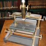
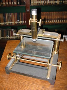
And lastly, one of the most unusual microscopes in our collection, a reversed microscope made by Nachet & Fils of Paris. The sample lies on the stage and can be illuminated from above, with the observer’s light path underneath the stage. It appears in the 1898 Nachet catalogue. This microscope was given to the collection by Dr and Mrs ten Hoopen, both of whom worked in the Department of Applied Botany before volunteering to help with the Archive and Museum after they retired.
The Delft School of Microbiology Archive owes a huge debt to both of them. Truus undertook two major tasks. First she restored our enormous collection of glass negatives (see the blogpost “Glass negatives galore!”) after they all got damp during asbestos removal in the attic where they were stored. She then went on to research and catalogue our collections of original and printed botanic and microbiological wall charts (see blogposts “The art of Henriette Beijerinck” and “Educational Wall Charts – where are they now?”). Meanwhile, Hens catalogued the microscopes and other equipment (we have the most amazing range of pH meters, for example) in our collection.
Without Truus and Hens ten Hoopen, we would know a lot less about the Collection than we do.

Reversed light path microscope by Nachet et fils
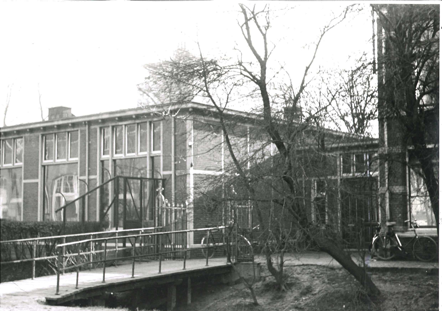
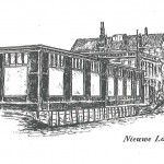
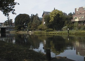
Delft University’s Biological Labs 1: The “Palace of Light” on the Nieuwelaan.
At the beginning of 2016, the Laboratory for Microbiology, together with the rest of the Department of Biotechnology, will move out of its current building, destination the new Faulty building on the outskirts of Delft. This seems like a good moment to take a look at the previous lab where so many breakthroughs were made. It was known as the “Palace of Light” because people worked late into the night.
We are fortunate that, thanks to a commemorative book published when Beijerinck was a fairly new Professor and photographs taken just before the department moved out, we know what it looked like. At the time it was built, it was the most expensive laboratory in the University. An extension was added in 1911.
Beijerinck’s laboratory
-
- The 1911 extension

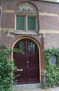
-
- The new door into the lab

Part of the building, on the left of the top photo, was the Professor’s house. The garden sloped gently down to a large canal (the canal’s edge can be seen as a white line across the bottom of the picture). It was in the greenhouse in this garden that the tobacco plants were grown for Beijerinck’s work on the Tobacco Mosaic Virus, and many famous microbial species were isolated from the soil and mud of the garden and canal.
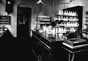
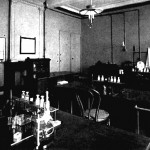
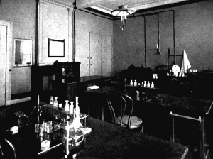
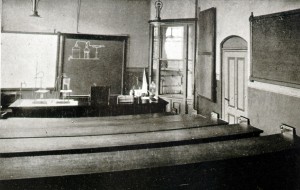
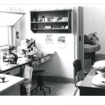
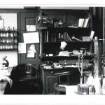
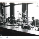
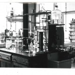
We have a few pictures of the inside of the lab in Beijerinck’s time.
-
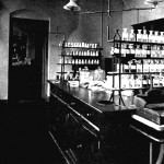
- staff lab
-
- student lab
-
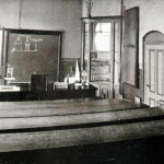
- lecture theatre
-
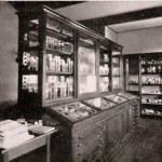
- culture collection
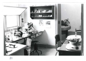
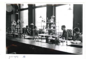
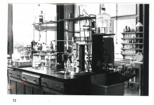
After Kluyver’s death and before everybody moved to the new (and current) building, everything was photographed. The photos are all numbered with corresponding floor plans so that we know not only which room is shown, but where the photographer was standing to take the picture. This is just a small sample.
-
- Kluyver's office as he left it.
-
- Kluyver's lecture theatre
-
- Electron microscopy
-
- Staff lab

-
- Students laboratory
-
- Laboratory
The building (without the extension) is still there. After a number of trials and tribulations including the building of a major bridge close to the front door of Beijerinck and Kluyver’s house, it is now apartments. To mark the 100th anniversary of Beijerinck’s appointment, a plaque was unveiled next to the original laboratory door.

The art of Henriette Beijerinck
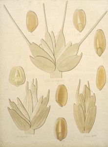
The laboratory was well supplied with printed botanic wall charts, but there was always a need for things that weren’t available. During Beijerinck’s professorship, his sister Henriette provided most of his display material, generally as large (A1 or A2 in modern terms) watercolours. Henriette Beijerinck was a qualified art teacher who presumably had private pupils, and in addition to material to illustrate lectures, also provided illustrations for several books.
-
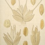
- agricultural grains
-
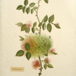
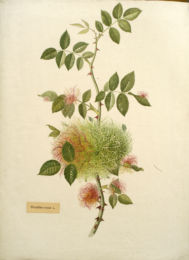
- rose gall
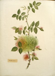
Professor Beijerinck’s earliest publications (while he was working at the Wageningen Agricultural Collece) were about the improvement of grains for agriculture, and our collection contains a number of drawings of seedheads. Since they are not signed, they could be by Henrietta, or even by Martinus. The image above left shows (clockwise from bottom left) wheat, European spelt and duram wheat. Beijerinck’s doctoral thesis (and a lifelong subject of research) was about plant galls and the collection includes quite a few pictures of galls. The illustration above right shows moss galls caused by Rhodites rosa gall wasp on young rose leaves.
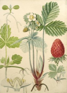
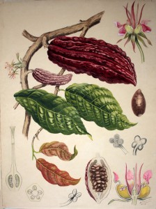
Henriette also painted assorted microorganisms (see, for examples, the blog post about possible life in comets), but her most beautiful works are the botanic charts. These three images are among the best.
-
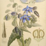
- Borage

-
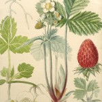
- Strawberry
-
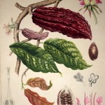
- Cocoa
Life at comet temperatures
With the increased interest in Comet 67P and ESA’s robot lander, Philae, this seems an opportune time to take a look at a small set of experiments carried out by Prof Beijerinck at the end of the 19th century. During the 1870s, there had been suggestions that life could have come to Earth from comets, but the discussion was largely theoretical until physicists found ways to replicate the temperatures found in space by liquefying gases.
In April 1907, Professor Heike Kamerlingh Onnes of Leiden University gave a demonstration of his new equipment for producing liquid gases (especially hydrogen) at the 11th Congress of the Holland Society of Sciences in Leiden. Beijerinck had been a member since the foundation of the Society in 1888 and it’s hard to imagine that he missed a chance to tour Kamerlingh Onnes’ brand new laboratory. It’s surely not a coincidence that in November and December of that year, he and his assistant, C.J. Jacobsen, took microorganisms from their collection to Leiden in order to test their ability to survive such extreme cold.
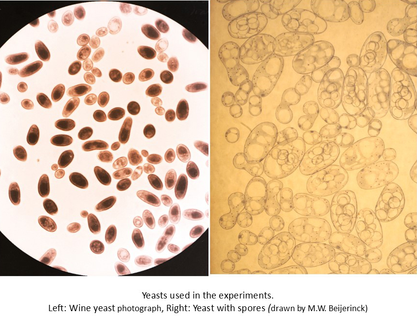
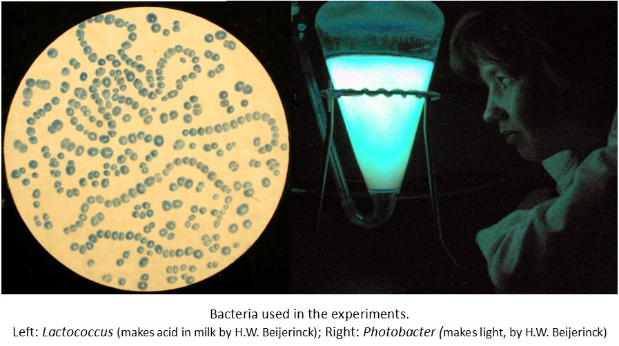
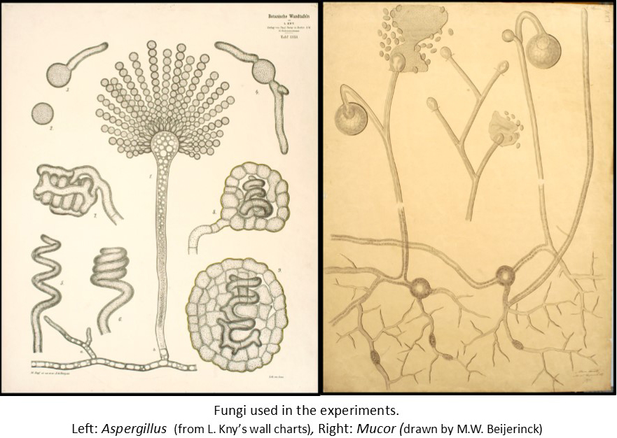
The experiments were very simple. They used a collection of microorganisms that they knew well, and whose behaviour under normal conditions they could predict. They chose bacteria that could make acid from milk and others that make their own light (bioluminescent), as well as cyanobacteria (also called blue-green algae). Among the “higher” microorganisms were yeasts that can make survival forms called spores, and others that can’t, as well as a couple of fungi which also make spores, and a green alga. Small amounts of each organism (in their growth medium) were sealed in small vials and then frozen in liquid nitrogen (N2; freezes at -195.8°C) or liquid hydrogen (H2; freezes at -253°C) for different lengths of time. The growth and behaviour of the microorganisms were then compared with cells that had not been frozen.
The first experiments used liquid N2 (probably because it was easier to produce) for 15 minutes. The second series used liquid N2 for 10 hours or liquid H2 for 45 minutes. The third series involved liquid N2 for 3 and 11 days. Finally, the microorganisms that had survived best were compared in liquid N2 over periods up to 15 days.
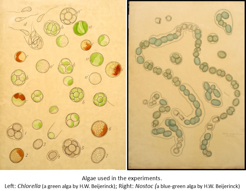
There was little difference between the N2 and H2 results – once the organisms were deep frozen, the extra drop in temperature made no obvious difference. The length of time frozen also made little difference. Survival varied with the microorganism involved. The spores of the fungi and yeast that could make them survived, but their active cells didn’t. The bacteria survived. The cyanobacteria all died and the higher green alga survived.
From these simple experiments, it could be concluded that simple microorganisms such as bacteria and organisms that make survival spores could survive in comets. Beijerinck also commented that extreme cold could not be used as a means of sterilisation.
Deep-freezing microorganisms appears to have been a curiosity for Beijerinck and he does not seem to have returned to the subject. We now know that more complex cells can survive if they are suspended in a solution with suitable protection, and they also do better if they’re frozen and thawed correctly. 100 years after Beijerinck’s experiments, deep freezing (usually in liquid N2) is now used to safely store all sorts of biological material from research stocks of bacteria and viruses to human sperm and tissues.
Recent Comments