
Posted in December 2017
Exploring the Delft School’s microscope collection – 1.
As I have mentioned before, the Delft School of Microbiology Archive has a small collection of old and unusual microscopes. We also have a range of attachments with varying (and sometimes unidentified) functions as well as two boxes of mysterious parts. One of my projects for this winter is to try as many of these things out as possible, and write a methods book for the collection.
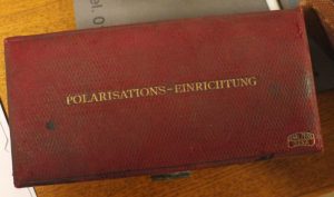
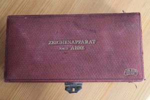
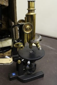
I have started with two red boxes from Ernst Abbe’s time at Carl Zeiss Jena (CZJ). Abbe was one of the founding fathers of CZJ and the inventor of many microscope techniques that we take for granted today, including standardizing lens quality. One of the boxes contains the parts necessary to convert a simple microscope for work with polarization, the other contains a camera lucida. Both were obviously routinely used in the times of Beijerinck, Van Iterson and Kluyver and we have enough of each for every student in a practical class to use them. None of the boxes included instructions, and nobody I’ve asked had tried to use either one. Since they turn up sometimes on auction sites, it seems to be worth outlining my findings here. I used my late 19th century jug-handled CZJ microscope.
-
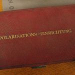
- Abbe's polarizer
-
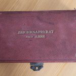
- Abbe's camera lucida
-
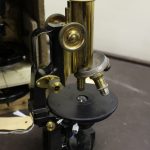
- My CZJ microscope
The polarizer.
The box contains a polarizer (top right in the photo), a calibrated ring (centre) and an eyepiece (bottom left). It took me a while to realize that the polarizer is actually two pieces which unscrew. The piece with the lens fits into the filter ring under the condenser, and the ring screws on underneath the filter ring to stabilize the polarizer. The calibrated ring fits around, and the eyepiece over the ocular.
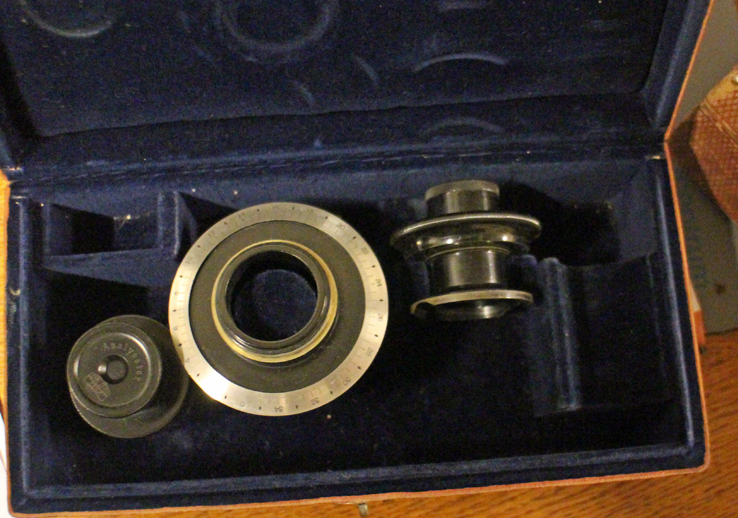
The contents of the polarizer box.
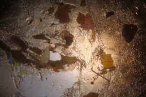
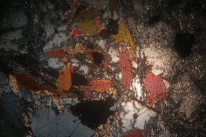
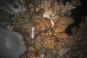
Using my usual LED lamp, I adjusted the mirror and condenser to give optimum light and then inserted a slide. Rather than microorganisms, for these experiments it was simpler to use samples guaranteed to give the dramatic colour changes associated with polarizing microscopy and so I chose mineral samples prepared for the microscope by Dr F Krantz of Bonn around the beginning of the 20th century (and also in our collection). The pairs of photos show the extremes of uncrossed and crossed polarized light paths for 3 different minerals.


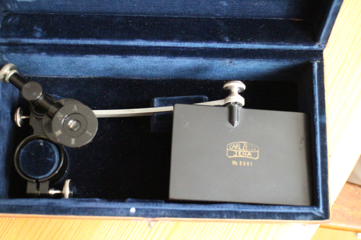
The camera lucida
The box contains a ring which fits over the microscope’s ocular. Attached to the ring is an arm with a mirror at its end and a shorter, moveable arm supporting the prism that combines the images from the microscope and the mirror. The arm allows the prism to swing over or away from the ocular, allowing the microscope to be used with and without it (very useful for focusing and sample placing).
Initially, I found this attachment very frustrating because everyone I had discussed it with confidently said that it projected the sample’s image onto paper beside the microscope. This did not happen. It was only when I looked through the ocular to check the microscope’s focus and saw a ghostly pen superimposed on the sample that I realised that the projection was the other way round! Thus far, I am not very satisfied with my photographs of the combined images, but I’ll post a picture here when I’m happy with them. The problem is not in using the equipment, but in convincing the camera that it can focus on the pen and sample at the same time – I see that CZJ’s catalogues of the time also offer a drawing platform for use with their camera lucida which was presumably exactly the correct height. If anyone wants to try, I’ve had better results with cream coloured paper rather than bright white paper which tends to reflect more in the field of view. Reducing the light in the room also helps.
Recent Comments