
Delft Microbiology
Life at comet temperatures
With the increased interest in Comet 67P and ESA’s robot lander, Philae, this seems an opportune time to take a look at a small set of experiments carried out by Prof Beijerinck at the end of the 19th century. During the 1870s, there had been suggestions that life could have come to Earth from comets, but the discussion was largely theoretical until physicists found ways to replicate the temperatures found in space by liquefying gases.
In April 1907, Professor Heike Kamerlingh Onnes of Leiden University gave a demonstration of his new equipment for producing liquid gases (especially hydrogen) at the 11th Congress of the Holland Society of Sciences in Leiden. Beijerinck had been a member since the foundation of the Society in 1888 and it’s hard to imagine that he missed a chance to tour Kamerlingh Onnes’ brand new laboratory. It’s surely not a coincidence that in November and December of that year, he and his assistant, C.J. Jacobsen, took microorganisms from their collection to Leiden in order to test their ability to survive such extreme cold.
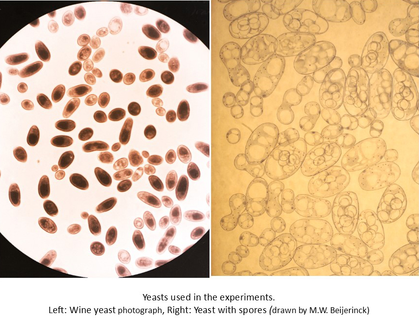
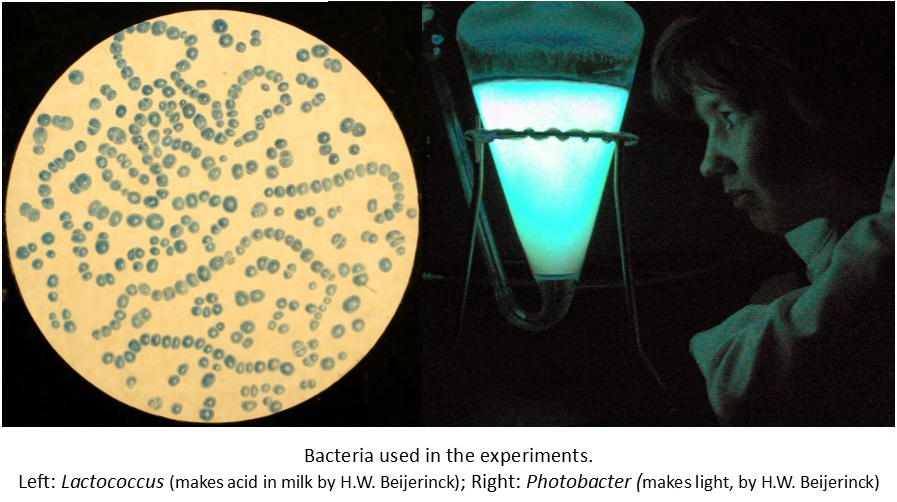
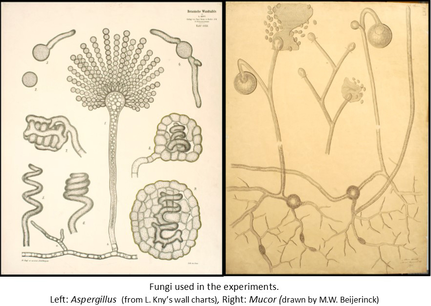
The experiments were very simple. They used a collection of microorganisms that they knew well, and whose behaviour under normal conditions they could predict. They chose bacteria that could make acid from milk and others that make their own light (bioluminescent), as well as cyanobacteria (also called blue-green algae). Among the “higher” microorganisms were yeasts that can make survival forms called spores, and others that can’t, as well as a couple of fungi which also make spores, and a green alga. Small amounts of each organism (in their growth medium) were sealed in small vials and then frozen in liquid nitrogen (N2; freezes at -195.8°C) or liquid hydrogen (H2; freezes at -253°C) for different lengths of time. The growth and behaviour of the microorganisms were then compared with cells that had not been frozen.
The first experiments used liquid N2 (probably because it was easier to produce) for 15 minutes. The second series used liquid N2 for 10 hours or liquid H2 for 45 minutes. The third series involved liquid N2 for 3 and 11 days. Finally, the microorganisms that had survived best were compared in liquid N2 over periods up to 15 days.
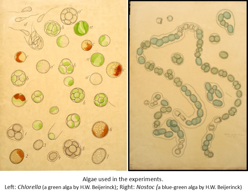
There was little difference between the N2 and H2 results – once the organisms were deep frozen, the extra drop in temperature made no obvious difference. The length of time frozen also made little difference. Survival varied with the microorganism involved. The spores of the fungi and yeast that could make them survived, but their active cells didn’t. The bacteria survived. The cyanobacteria all died and the higher green alga survived.
From these simple experiments, it could be concluded that simple microorganisms such as bacteria and organisms that make survival spores could survive in comets. Beijerinck also commented that extreme cold could not be used as a means of sterilisation.
Deep-freezing microorganisms appears to have been a curiosity for Beijerinck and he does not seem to have returned to the subject. We now know that more complex cells can survive if they are suspended in a solution with suitable protection, and they also do better if they’re frozen and thawed correctly. 100 years after Beijerinck’s experiments, deep freezing (usually in liquid N2) is now used to safely store all sorts of biological material from research stocks of bacteria and viruses to human sperm and tissues.
Celebrating Professors
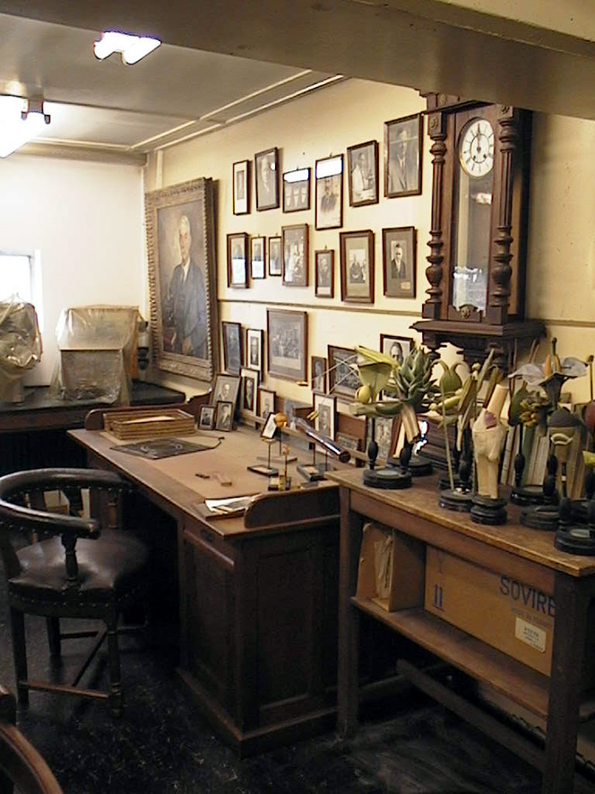
During the first half of the 20th century, it seems to have been customary to make certificates, posters or books to commemorate special events. The Archive includes a number of photo albums showing laboratories in the University or even abroad, but three examples stand out, not least because of the considerable amount of work that went into them.
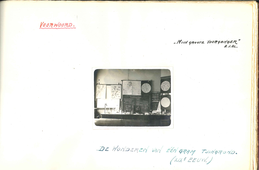
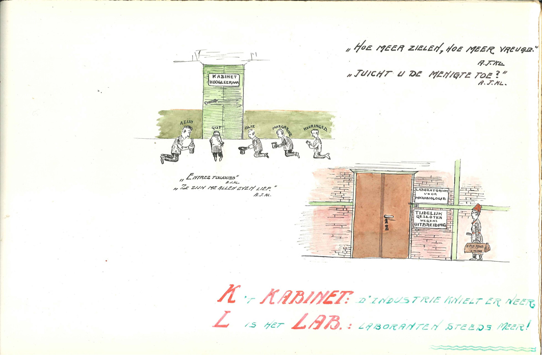
The first is a book made for Kluyver by three of his pupils (van Niel, Leeflang and Struyk) a few years after he became Delft’s Professor of Microbiology. The book compares Beijerinck’s 19th century approach to the wonders to be found in 1 gram of soil with Kluyver’s 20th century approach to the wonders associated with 1 gram of carbinol. That’s not students kneeling outside the Professor’s door in the 4th page, but representatives of industry!


The second is a handmade poster (about the size of a large double bed) that was made to mark the 25th anniversary of Kluyver’s inaugural lecture. It shows notable features from those 25 years, including sketches of the laboratory, Kluyver’s most famous work (The Unity in Diversity) and his inaugural address (“Rede”) in which he emphasized the importance of applied research. Every rectangle represents a story.
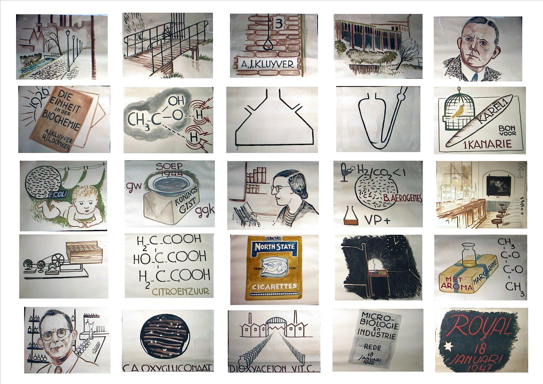
The Kluyver Flask (still used for growing submerged, well-mixed cultures) and a shaker for closed jars containing oxygen-free cultures are shown. During the Dutch “Starvation Winter” at the end of World War II, Delft’s Yeast and Spirits Factory gave their staff soup made from yeast extract at lunch time, and as one of their advisors, Kluyver regularly benefited from this at weekly meetings. Lastly, at a time when it was usual to stand if a Professor came into the room, the staff’s affection for Kluyver shines through in several squares teasing him about his smoking!
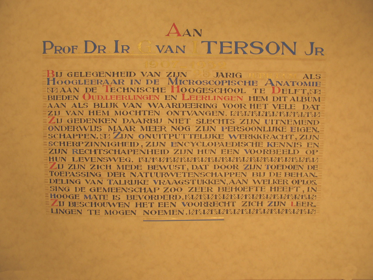
A book to mark van Iterson’s 25th anniversary as a Professor also falls into this category despite being essentially a photo album because the makers included all of his PhD students, postdocs and co-workers from other countries, showing who they were, what they did and what happened to them afterwards. van Iterson’s conviction that the primary job of a Professor is to teach is obvious from the fact that there’s 160 pages, each with one or more person on it. The example here shows J.E. van Amstel, the first woman to be granted a Doctorate in Delft.

Here there be surprises!
Thanks to the hard work of volunteers over the last couple of decades, the catalogues for the various collections in the Archive are gradually becoming more detailed, and this continues, in combination with document scanning. Also, now that we’re going to be moving to another building at the end of the year, the current team has been investigating cupboards and boxes that were fairly low down the priority list. We’re often surprised by what we find. Many things shed light on how things were done in the late 19th and early 20th centuries, others are simply curiosities which may add to our questions even as they answer others. For example, letters by van Iterson to an instrument maker explained the envelope full of paper discs labelled “anorthoscope” and we then discovered in the Instrument Catalogue that we still have one! Anorthoscopes might be considered to be a forerunner of movies where the user turned a handle and figures on paper discs seemed to walk or run. Judging by the results of internet searches, they’re largely considered to be toys, but van Iterson was obviously using them for some sort of research, although we still aren’t sure what! The images with this post show examples of other finds.
In roughly chronological order:
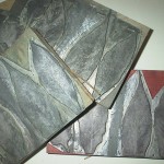
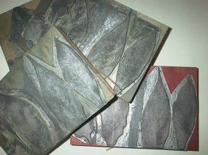
Beijerinck started as a botanist (the discipline of microbiology hadn’t really been invented then) and wrote his PhD thesis on plant galls. It was obviously a subject close to his heart since he kept his samples, including a couple of boxes containing 19th century pillboxes full of different sorts of gall.
Beijerinck’s work on TMV is credited as being the first to establish that the causative effect of Tobacco Mosaic Disease was self-replicating and thus not a chemical. That we have his lab journals is well-known, but we also have the plates used to illustrate that first paper (and many others). There’s actually 5 plates in this set, each slightly different so that the published image was correctly coloured.
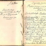
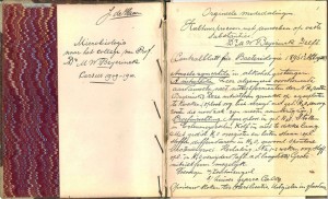
A fairly recent gift to the collection was a set of brown notebooks that were found after the death of the donor’s relative. These proved to be a full set of lecture notes made by a student during Beijerinck’s microbiology lecture series. We’ve also been given the set of notes that Kluyver made for his speeches when his PhD students were awarded their Doctorates, among other things. I love the moment when someone comes through the door and says “someone I know is clearing a house and wants to know whether the museum wants this”…
Harry Barker spent a year with Kluyver in the 1930s, doing some of his early work on methane-producing bacteria, and his slides are among a pile of microscope preparations from that period that turned up in a cupboard. As well as the slides, we have glass bottles containing chemicals, preparations and samples, including a very early enzyme extract labelled “lactase”. There was still measurable activity in the sample when it was tested in 1989 despite the fact that storage conditions have been less than optimal over the century since it was made.
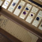
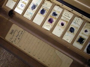
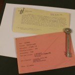
During WW2, Kluyver was Chairman of a committee that tried to maintain contact with the students who’d been taken as forced labour to work in Germany. We still have many of the papers relating to that time, including the card indexes showing where the students were, and where they were working, as well as correspondence from students and the authorities. (Ignore the key, it’s just there to hold the papers flat.)
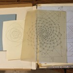
When Mrs van Iterson visited the laboratory (quite a long time ago), she commented (more than once) that she wished that her husband had had a computer. When one looks at the drawings and calculations (made with nothing more than graph paper, log tables and a slide rule) it’s easy to see why. His work on phyllotaxis is still regarded as relevant today, and is a tribute to the days and days of patient calculation and drawing that formed the foundations of modern mathematical modelling.
-
- Beijerinck's dried plant galls
-
- Notes from Beijerinck lectures
-
- Printing plates
-
- Barker's microscope slides
-
- WW2 student information

-
- van Iterson's drawings

Educational wall charts – where are they now?
During the second half of the 19th century and the early years of the 20th, a number of companies produced wall charts as teaching aids. The theme and quality varied enormously, but most of the ones intended for bioscience education are not only very detailed, but generally beautiful in their own right. They were sold singly, or by subscription. Subscribers were sent the charts as they became available (often 1-2 per year), together with explanatory books.
Delft’s collection includes several complete series, including those by Kny, Dodel-Port and the series known as the Tabulae Botanicae (often attributed to “Blakeslee et al”, but most of the posters are signed by R. Erlich). However, we also have a number of incomplete series which might be represented by a single example, or a few posters. Some complete series are available elsewhere. For example, the conifers chart shown here is number 16 of 50 by Albert Peter – a complete series is held by the University of Bourgogne. However, many seem to have been forgotten.
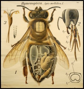
With the help of collections around the world, it has recently been possible to assemble an electronic complete series of Pfurscheller’s zoological charts (here represented by the fly). Representatives from other partial series are shown here:
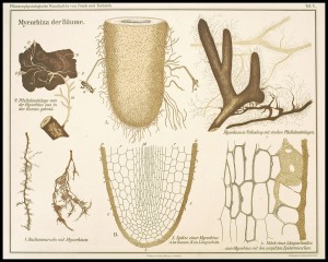
The Mycorhiza chart is number 10 from “Pflanzenphysiolgische Wandtafeln” by Frank & Tschirch (we have 1-10 of 60), most of the series is held by the University of Utrecht, among others.
The sweetcorn is number 3 in series C of a set for general biology by Haecker & Mülberger. Series A and B are both currently known by single examples, and Delft currently has 1-4 of series C, the size of the complete set is unknown.
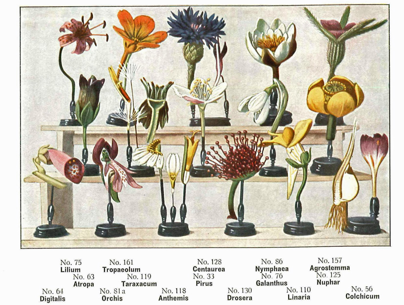
The flowers come from a set by O.W. Thome (Delft has numbers 15 & 23).
The microorganisms come from a series by W. Henneberg about microorganisms with positive or negative impacts on the fermentation industry. This is number 6, vinegar fermentation. Delft has 8 of an unknown number.


Glass negatives galore!

Small projector set up for glass negatives.
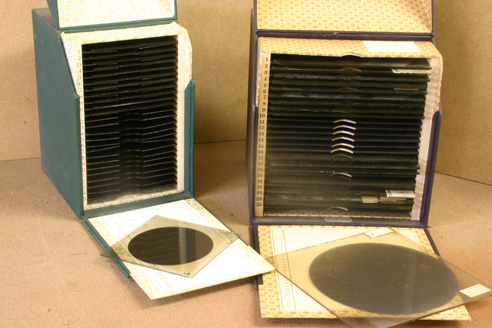
Glass negatives.
Our glass negative collection (about 27,000 of them) dates from the mid-1880s to the 1960s (when they were being used with the electron microscope. It covers a remarkably wide range of topics including Beijerinck’s gall wasps, light microscopy, travel (especially van Iterson’s working trip to Indonesia) to images from publications – essentially what any group of Professors would have in their Powerpoint collections today. The quality of the images is amazing – it has been possible to enlarge pictures to A0 without grain or blurring.
Later posts will showcase the individual collections of the three Professors, this is just a taster to show examples of what we have.


Delft’s first microbiologist – Antonie van Leeuwenhoek
Although the Delft School of Microbiology only dates back to Martinus Beijerinck and the late 19th century, it seems churlish to ignore Antonie van Leeuwenhoek on a blog discussing Delft microbiology just because he was 200 years too early. He was not a teacher and indeed actively resisted explaining his methods, but he did publish copiously about everything he saw with his magnifying glasses and simple microscopes, making him the first microbiologist (although not the first microscopist).
Today, van Leeuwenhoek is generally mentioned in connection with the discovery of microorganisms. However, his studies were much broader than that. He dissected insects, and examined anything that would fit on his microscope. His first letter to the Royal Society illustrates this clearly as it covers the sting, head and eye of the bee, and the structure of a louse as well as his observations of fungus that he said grew on leather, meat and other things.
Van Leeuwenhoek’s microbiological discoveries began in 1674 when he examined samples from the cloudy water of the Berkelsemeer, a lake near Delft that no longer exists, and found his famous “little animals”. His discovery of bacteria probably dates from his pepper water experiments in 1676, when he reported seeing extremely small animals among the others – a copy of the drawing that accompanied this letter was published by Henry Baker, and is shown here. “Fig IV” is probably the first appearance in print of a bacterium.
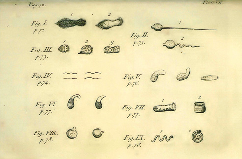
Baker’s copy of AvL’s pepper water illustration.
-
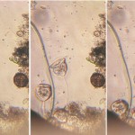
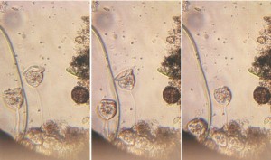
- Bright field microscopy of living protozoa
-
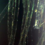
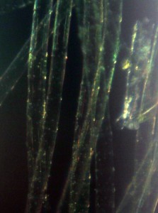
- Dark field microscopy of algae
-
- Facsimiles of van Leeuwenhoek microscopes
The film clip here – www.youtube.com/watch?v=OniSF8QrHac – shows what can be seen with facsimiles of van Leeuwenhoek microscopes.
And there’s an excellent website about our Founding Father here: http://lensonleeuwenhoek.net/
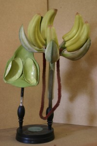
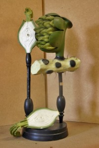
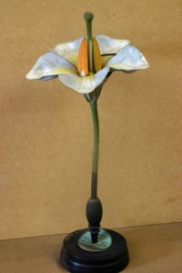
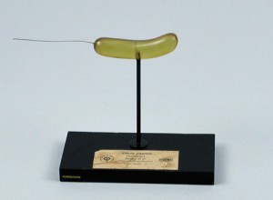
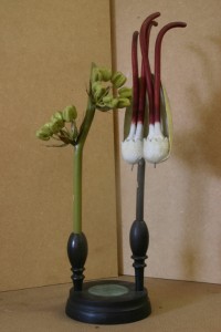
Brendel flower models
The collection includes about 20 of the flower models made by Robert and Reinhold Brendel in the late 19th and early 20th centuries. The models are made from papier-mâché with other materials including plaster, glass beads, wood, cotton and rattan added to give detail or texture. They can be taken apart to reveal internal details (although they’re not always simple to put back together again). The Brendels were advised on the accuracy of their models by various Professors, depending on what the particular model was intended to show. As well as plants, models of fungi, yeast and bacteria were eventually included.
Dealers included the models in their own catalogues, but Delft has the only known surviving Brendel catalogue, issued in Berlin in 1913.

The Delft School of Microbiology Archives
The “Delft School of Microbiology” is based on the work of our first three biological Professors – Beijerinck, van Iterson and Kluyver. The Archive is a collection of their papers, teaching aids, furniture and other materials and functions as a small museum in the Department of Biotechnology.
At the moment, the collection is being digitised and catalogued. This blog will report on some of the interesting and sometimes unexpected things to be found in the collection.
We’ll also cast an occaisional glance at our honorary member, the original Delft microscopist and microbiologist, Antonie van Leeuwenhoek.
Recent Comments