
Posted in 2018
We’re open on Sunday 28th October!
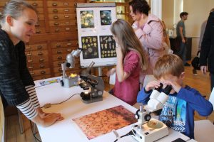
The Delft Science Centre will, like many other places, be open for “Wetenschapsdag” (Science Day) on 28 October 2018 between 11:00-16:00.
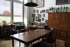
Among the exhibits, we will be showing what you can do with microscopes old and new, and the Beijerinck Office Museum will be open to escorted groups.
The Minerals Museum will also be open, with VR demonstrations.

The full programme (in Dutch) is available here: https://www.tudelft.nl/sciencecentre/activiteiten/wetenschapsdag/.
Presenting historical microscopes
Now that we’ve finished unpacking and sorting the collection, and the cataloging is well under way, the next priority is to improve the way that it is presented to the public. In this context, “public” can mean anyone from an international group of Professors on a visit to the Netherlands to members of the general public who visit the Science Centre at the weekend. In anything that we do, we must reach a happy balance between too much or too little explanation, without cluttering up any displays.
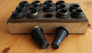
We also have the interesting problem of several boxes (labelled “microscope parts”) of mysterious lenses from unknown makers and other items. Fortunately, we have now teamed up with the Dutch Foundation for Historical Microscopy (here: http://stichtinghistorischemicroscopie.nl/en/) to try and solve the mysteries.
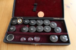
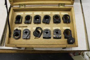
Some of the stars of our Brendel flower model and microscope collections can now be seen in display cases in the public areas of the Centre, but we have much more material than we can display. Some of the brown wooden boxes contain equipment such as microtomes, microforges and densitometers, so it was a useful exercise to investigate them all.
For good cataloging as well as publicity, everything needed to be well photographed. Jan Sluijter, the professional photographer who also worked on the Science Centre’s Minerals Collection (which goes back to the middle of the 19th century), therefore spent a fair amount of the winter/spring here.
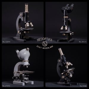
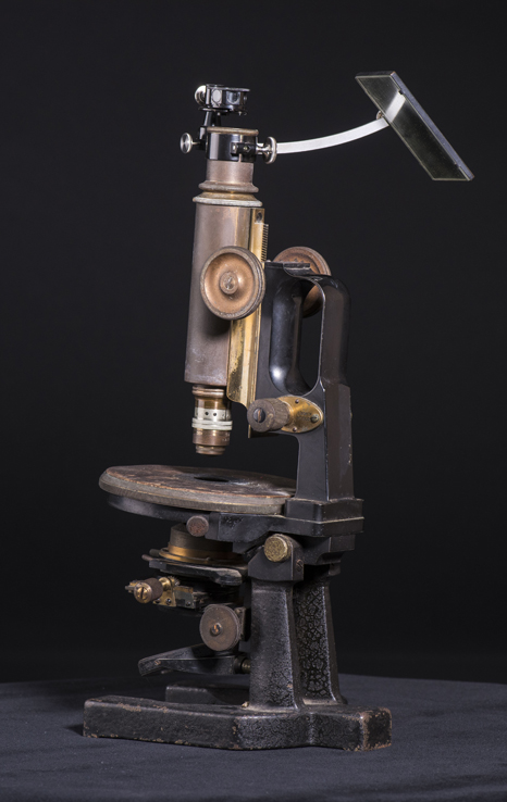
We decided that we did not want to try and restore any items to factory perfection, but to leave the evidence of their long working lives. Some microscopes had obviously only had one careful owner (to borrow from car salesmen), others had been used by hundres of students in practicals. In addition, Jan photographed everything on both black and white backgrounds and made short film clips showing each item rotating on a turntable. It’s amazing how much difference the colour of the background makes, as can be seen in the two photos of the same Carl Zeiss Jena “jug handled” microscope with fitted camera lucida, below.
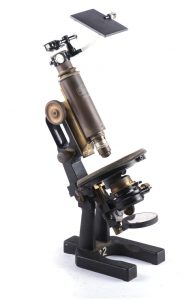
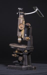
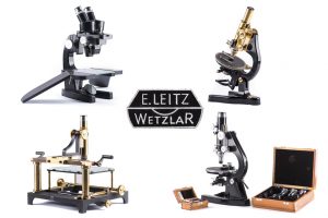
Jan’s now experimenting with different ways of showing representativeparts of the collection (see the Leitz and Bleeker microscopes, below). I still can’t decide whether I prefer the white or black backgrounds, but I think that the black is winning!
Delft’s Science Centre nominated for Museum Prize!
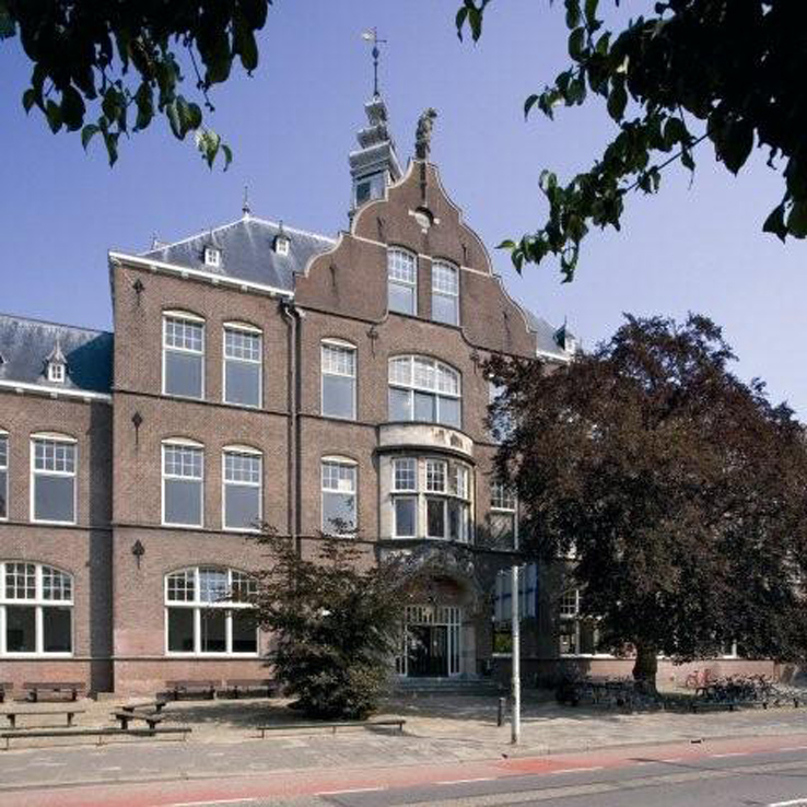
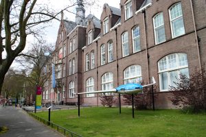
Delft Science Centre with new Kamer van Beijerinck, 2nd floor with balcony
We’re very excited to be able to tell you that the Delft Science Centre, home of the Beijerinck, van Iterson and Kluyver collection as well as the Faculty of Mining’s mineral collection and more modern exhibits from Delft University of Technology, has been nominated for the BankGiro Lottery’s Museum Prize.
It would be very kind and much appreciated if you would support us by voting.
The voting site is in Dutch, so here’s a translation into English for those who use other languages.
1; Go to the site here:
https://www.museumprijs.nl/genomineerden/science-centre-delft
2; Hit the green button labelled “Stem op dit museum”.
3: When the blue rectangle comes up, it says “Yes, I’m voting for the Science Centre Delft” and asks you to fill in your name and then your email address (that’s so that they can make sure that each email address is linked to one vote and so that they can let you know if you win the prize trip to New York).
Also click the box immediately under the email box – it indicates that you’re over 18 and agree with the rules (which you can see by hitting the “actievoorwaarden” link. If you want to read them, copy the text and paste it into Google Translate or another translation app). The two boxes below, if clicked, let them send you news about the BankGiro Lottery (who’re sponsoring the prize) and the Prins Bernhard Culture Fund (also sponsors).
4: Click on the green button.
You will then be thanked for voting and they will send an email to the address you gave them. The email will ask you to click on another green button to confirm your vote (it prevents folk from getting carried away and making up email addresses to let them vote often).
Thoughts on Van Leeuwenhoek’s optical tool box
My personal project to find the descriptions of methods buried in Van Leeuwenhoek’s letters and to try and work out how he did things that he didn’t describe continues. My most recent results have now appeared in the Nov 2017 FEMS Letters with the POI: https://doi.org/10.1093/femsle/fnx247. The paper covers problems encountered with using the classical microscopes with some samples that we know he studied, and suggests a solution.
Antoni van Leeuwenhoek did not limit his research to microorganisms. Indeed, he worked with a very wide range of samples ranging from wood and animal tissues through insects to individual cells and sperm to bacteria. His microscopes work very well with transparent samples where the light can pass through them (and the samples were small) but, as can be seen from Figure 1, he was obviously also using reflected light.
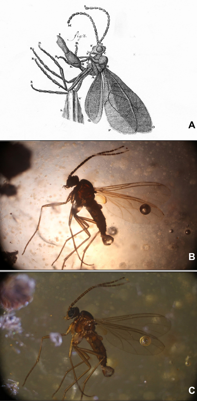
Transmitted and reflected lighting of a gnat
Figure 1A shows a drawing of a gnat, taken from a 1702 Van Leeuwenhoek letter. Figures 1B and C. show a similar insect photographed under a 19th century Carl Zeiss Jena microscope with transmitted (B) and reflected (C) light. It is clear that the level of detail shown by Van Leeuwenhoek cannot be observed on the silhouette shown in B.
The details are all in my papers, but my conclusion is that, as he did with his aalkijkers (his microscopes for viewing the blood in the capillaries of the tails of small eels and fish where the sample pin was replaced by a glass tube to keep the specimen alive), Van Leeuwenhoek adapted his magnifiers to suit the task in hand.
Figure 2 shows a collection of magnifying equipment that I think Van Leeuwenhoek used as his optical tookbox. The photographs show modern facsimiles, the drawings were made during or just after Van Leeuwenhoek’s life. This conclusion is based on experimental results and historical documents.
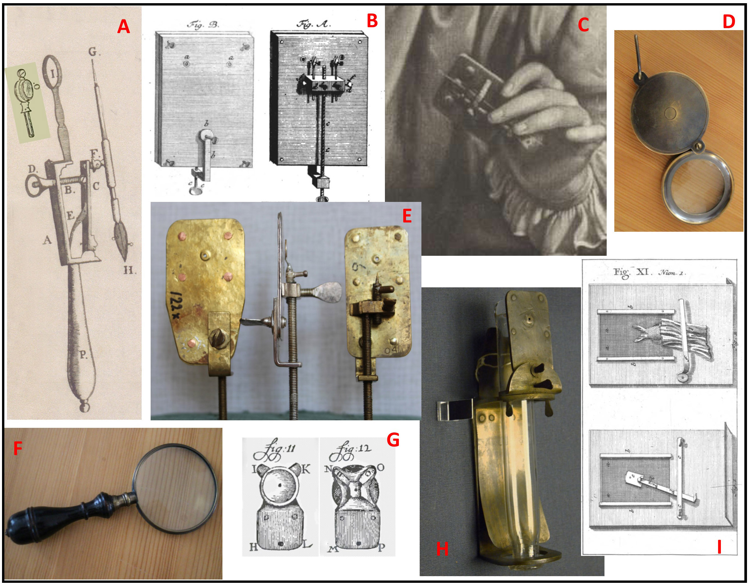
Figure 2: Optical toolbox
A: This magnifying glass (or something very like it) appears in the frontispiece of the auction catalogue for the sale of Van Leeuwenhoek’s microscopes after his daughter’s death. Because the lens is set into a simple ring rather than the metal plate of the Van Leeuwenhoek microscope, the setting does not cast a shadow on the sample if reflected lighting is used.
B: This version of the Van Leeuwenhoek microscope was published by a German visitor (Zacharius-Conrad von Uffenbach), and microscopes with 2 lenses are mentioned in the sale catalogue. With 2 different lenses side by side, it would have been more convenient than moving a fragile sample to another microscope for a different magnification.
C: This 3-lensed microscope comes from an engraving made while Van Leeuwenhoek was in the prime of life, and is also mentioned in the catalogue. Note the holder for a capillary (used for liquid samples such as blood) beside the sample needle.
D and F: Copies of 17/18th century magnifying glasses, as sold by the Rijksmuseum Boerhaave. “Burning glasses” are mentioned in the inventory of the Van Leeuwenhoek house after Maria van Leeuwenhoek’s death. As well as magnifying larger samples, they can be used to control the lighting of samples on the microscopes.
E: Some of the facsimiles of existing Van Leeuwenhoek microscopes made by Hans Loncke and used in my experiments.
G: Van Leeuwenhoek’s adaption of the eyepiece on his aalkijker to allow reflected lighting while protecting the eye with a small cup.
H: A facsimile of the aalkijker drawn in Van Leeuwenhoek’s first paper on blood circulation. Note the use of the same lens plate as with the microscopes.
I: His final adaption of the aalkijker to make its use easier for visitors, as mentioned by him in a letter and drawn by Von Uffenbach.
People often describe his microscopes as crude, and perhaps they are when compared to other, ornamented microscopes of his time, but I suspect that Antoni van Leeuwenhoek was more interested in making something that would allow him to carry out his experiments rather than producing elegant instruments.
Recent Comments